MP Board Class 12th Biology Important Questions Chapter 3 Human Reproduction
Human Reproduction Important Questions
Human Reproduction Objective Type Questions
Question 1.
Choose the correct answers:
Question 1.
The number of chromosome in human cell will be :
(a) 23
(b) 46
(c) 92
(d) 22
Answer:
(b) 46
Question 2.
The hormone estrogen is secreted by :
(a) Corpus luteum
(b) Graafian follicle
(c) Uterus
(d) Vagina.
Answer:
(b) Graafian follicle
Question 3.
The copulatory organ in female human being is :
(a) Fallopian tube
(b) Uterus
(c) Clitoris
(d) Vagina.
Answer:
(c) Clitoris
Question 4.
The gestation period in rabbit is about :
(a) 27 days
(b) 22 – 30 days
(c) 45 days
(d) 28 days.
Answer:
(b) 22 – 30 days
![]()
Question 5.
Hormone secreted during delivery is:
(a) Progesterone
(b) Thyroxine
(c) Relaxin
(d) Glucocorticoid.
Answer:
(c) Relaxin
Question 6.
Blastopore is formed in :
(a) Gastrula
(b) Blastula
(c) Morula
(d) Nurmula
Answer:
(a) Gastrula
Question 7.
Number of parents involved in asexual reproduction is :
(a) Many
(b) Three
(c) Two
(d) One
Answer:
(d) One
Question 8.
Sertoli cells are found in :
(a) Kidney
(b) Ovary
(c) Liver
(d) Testis.
Answer:
(d) Testis.
Question 9.
Which two are found in human semen in more quantity :
(a) Ribos and potassium
(b) Fructose and calcium
(c) Glucose and calcium
(d) DNA and testosteron.
Answer:
(b) Fructose and calcium
Question 10.
Which part of the fallopian tube is nearest to ovary :
(a) Ampula
(b) Isthmus
(c) Infundibulum
(d) DNA and testosteron.
Answer:
(c) Infundibulum
Question 11.
In ovarian cycle of human ovulation is starts from :
(a) 1st day
(b) 5th day
(c) 14th day
(d) 28th day.
Answer:
(c) 14th day
Question 12.
Which hormone controls the sperm formation :
(a) ADH
(b) FSH
(c) LH
(d) STH.
Answer:
(b) FSH
Question 13.
Which is responsible for movement of sperm :
(a) Cilia
(b) Flagella
(c) Basal body
(d) Nucleosome.
Answer:
(b) Flagella
![]()
Question 14.
Sperms are mature in :
(a) In oviduct
(b) In epididymis
(c) In vagina
(d) All of the above
Answer:
(b) In epididymis
Question 15.
Seminiferous tubules are found in :
(a) In testis
(b) In ovary
(c) In kidney
(d) In lungs.
Answer:
(a) In testis
Question 2.
Fill in the blanks :
- The nature of semen is ………………
- The male sex hormone testosterone is produced by ……………… in testis.
- The structure between placenta and foetus is called ………………
- Human being are ……………… breeders.
- The site of fertilization and implantation in human female is ……………… and ………………
- After ……………… year growth does not occur in girls.
- ……………… hormone is secreted during child birth.
- Segments of testis is consist of ……………… tubules.
- Testis starts the secretion of ……………… hormone at puberty.
- The process of fertilization is occurs in ……………… tube.
- ……………… provides the nutrition to embryo.
- ……………… hormones are secreted by graafian follicle.
Answer:
- Alkaline
- Leyding cells
- Umblical cord
- Continuous
- Oviducts, uterus
- 14
- Relaxin
- Seminiferous tubules
- Testosteron
- Follopean tube
- Placenta
- Estrogen and Progesteron.
Question 3.
Match the followings:
I.
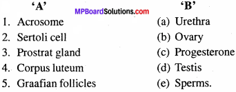
Answer:
- (e)
- (d)
- (a)
- (c)
- (b).
II.
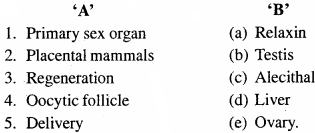
Answer:
- (b)
- (c)
- (d)
- (e)
- (a).
Question 4.
Write the answer in one word/sentences:
- How many sperms will be produced from 24 spermatocytes during spermatogenesis?
- Where does fertilization takes place in mammals?
- How many polar bodies are formed during the formation of one gamete?
- How many polar bodies are formed during the formation of ovum during oogenesis?
- Write the name of three layers of gastrula.
- Spermatogenesis is the example of.
- How many germ layers are found in gastrula stage?
- Write the duration of gastation period in human cases.
- How many autosomes are found in human sperm?
- Name the process in which formation and maturation of ova.
- Name the main store house of sperms.
- Skeletal muscle is originated by these layer.
- Name the process in which zygote starts dividing by specific mitotic divisions.
- In which month body hair developed in embryo.
Answer:
- 1.96
- Fallopian tube
- 2
- 3
- Ectoderm, Mesoderm and Endoderm
- Male gamete formation
- Three
- 280 days
- 22
- Oogenesis
- Epididymis
- Ectoderm
- Cleavage
- Fourth month.
Human Reproduction Very Short Answer Type Questions
Question 1.
After fertilization, how many days are needed for the birth of human child?
Answer:
280 days (9 months and 10 days).
Question 2.
To produce the new organisms of their own kind is called by a term, name it.
Answer:
Reproduction.
Question 3.
By which name, the reproduction happens by means of fusion of two different gametes?
Answer:
Sexual reproduction.
![]()
Question 4.
Which gametogenesis goes on till life span in human beings after attaining puberty?
Answer:
Spermatogenesis.
Question 5.
The stoppage of menstrual cycle in a 50 yrs. old female is known as.
Answer:
Menopause.
Question 6.
What is pregnancy?
Answer: The time period between fertilization to the birth of child is called pregnancy.
Question 7.
Where is carpora cavernosa found?
Answer:
Carpora cavernosa is found in penis.
Question 8.
What is the gestation period in elephant, dog and cat?
Answer:
Elephant 641 days, dog 58 – 68 days, cat 63 days.
Question 9.
Name the hormone that relaxes pubic symphysis during parturition.
Answer:
Relaxin hormone.
Human Reproduction Short Answer Type Questions
Question 1.
Why testes are found outside the abdominal cavity in males?
Answer:
In male, one pair of testes are found in the scrotum or scrotal sac, which are situated outside the abdomen. Temperature of this scrotum is always 2°C less than the body temperature, which is suitable for formation and growth of sperm, otherwise spermatogenesis will not occur. Therefore, testes are situated outside the abdomen in man.
Question 2.
What will happen if leydig cells of testis in males are destroyed?
Answer:
The endocrine cells of the testis are the leydig cells, they are situated in between the seminiferous tubules. Leydig cells secrete the male sex hormones androgens. Testes secrete four types of androgens:
- Testosterone
- Androsterone
- Epiandosterone and
- Dihydroepiandrosterone.
Out of these male sex hormone is testosterone.
Functions of Testosterone:
- Testosterone stimulates testes to descend into the scrotum in embryos.
- It helps in the development of secondary sexual characters.
- Stimulates spermatogenesis.
If leydig cells are removed from the body of males, then the above mentioned functions will stop.
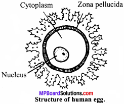
Question 3.
Describe the structure of human egg.
Or
Draw neat well – labelled diagram of human ovum which is ready for fertilization.
Answer:
The egg of human being is rounded and non – motile. It lacks yolk and contains a large Cytoplasm Zona pellucida. amount of cytoplasm. A nucleus (haploid) is present in the centre of egg. Whole egg is covered by a translucent and non – cellular membrane known as zona pellucida. The zona pellucida is irregularly covered by follicle cells. This layer is called as corona radiata. The cells of corona radiata are disintegrated before fertilization.
Question 4.
What is menstrual cycle ? Which hormones regulate menstrual cycle?
Answer:
The reproductive cycle in the female primates is called menstrual cycle. The uterus linings becomes thick and spongy to receive fertilised egg. If the egg is not fertilised, this lining is not needed any longer so, it slowly breaks and comes out through vagina along with blood and mucus. This is called menstruation. It is repeated at an average interval of about 28/29 days.
Following hormones regulate this cycle:
- Gonadotropin
- Estrogen
- Luteinizing hormone
- Follicular stimulating hormone
- Progesterone
![]()
Question 5.
What is parturition? Which hormones are involved in induction of parturition?
Answer:
The process of delivery of the foetus (child birth) at the end of the pregnancy is called parturition. The signals for parturition originate from the fully developed foetus and the placenta which trigger the release of oxytocin from the maternal pitutary. Oxytocin acts on the uterine muscles and induces stronger uterine contractions leading to expulsion of the baby. Relaxin hormone released by the ovary widens the vagina to facilitate birth.
Following hormones are involved in induction of parturition:
- Cortisol
- Estrogen
- Oxytocin.
Question 6.
In our society the women are often blamed for giving birth to daughters. Can you explain why this is not correct?
Answer:
Women are blamed for giving birth to daughters. This is wrong because sex of the baby is determined by the sperm that can have either X or Y – chromosome. Women have only one type of chromosome (X) in all the ova.
- If the sperm having X-chromosome fertillises the ovum (X), the resulting zygote (XX) will become a female.
- If the sperm having Y-chromosome fertillises the ovum (X), the resulting zygote (XY) will become a male.
Question 7.
How many eggs are released by a human ovary in a month? How many eggs do you think would have been released if the mother gave birth to identical twins? Would your answer change if the twins born were fraternal?
Answer:
Only one egg is released by a human (female) ovary in a month. Only one egg is released if the mother gave birth to identical twins. Yes, two or more eggs are released in case fraternal twins are born.
Question 8.
Name the hormones involved in regulation of spermatogenesis.
Answer:
The hormones involved in regulation of spermatogenesis are:
- Gonadotropin releasing hormone
- Luteinizing hormone (LH)
- Follicle stimulating hormone and
- Testosterone.
Question 9.
Define spermiogenesis and spermiation.
Answer:
Spermiogenesis:
The process involving transformation of spermatid into spermatozoa is called spermiogenesis.
Spermiation:
After spermiogenesis, sperm heads became embedded in the sertoli cells and are finally released from the seminiferous tubules by the process called spermiation.
Question 10.
Give composition of seminal plasma.
Answer:
Composition of seminal plasma : Fructose, calcium ion, some enzymes prostaglandins.
![]()
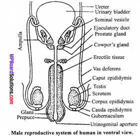
Question 11.
Draw a labelled diagram of male reproductive system.
Answer:
Question 12.
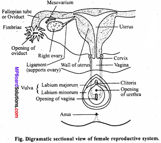
Draw a labelled diagram of female reproductive system.
Answer:
Question 13.
Write two major functions each of testis and ovary.
Answer:
Functions of Testis:
- Production of sperms by seminiferous tubules.
- Production of male sex hormone, testosterone by leydig cells.
Functions of Ovary:
- Production of ova (eggs).
- Production of female sex hormones, estrogen and progesterone.
Question 14.
Explain menarchy or menopause.
Answer:
Menopause:
In every human female, puberty period starts from 12 – 13 yrs of age to 45 – 50 yrs of the age. During this period except pregnancy at every interval of a month during 26th day to 28th day. If pregnancy does not occur then the internal wall of the uterus secretes out mucilaginous liquid along with blood. Secretion continues for 3 – 4 days called menses. As it comes at definite period so, it is called menstruation cycle. At the age of 40 – 50 yrs. menses stop and females reach to a stage called menopause. Ability of pregnancy also stops after attaining menopause.
Question 15.
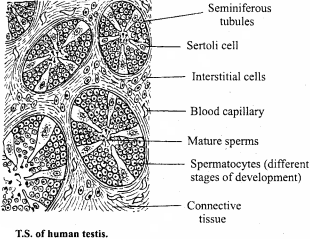
Draw a well labelled diagram of the T.S. of human testis.
Answer:
Question 16.
What is the position of fallopian tubule in female reproductive organs? What are their significance?
Answer:
Fallopian tubules are a pair of small muscular tubes, one each on either side of the uterus. These tubules extend from the vicinity of the ovary to the ovary. Each tubule is about 10 cm in length. The free end of each tube lies near the ovary of its side. This end is funnel shaped and fimbriated. It is called ostium and infundibulum. Infundibulum opens in the abdominal cavity by means of abdominal ostium. The fallopian tubule is kept in position by a mesentery which is attached to the uterus.
Significance or Functions of Fallopian Tubes:
- By their lashing movement of the cilia present in the lining of infundibulum and nearby area help in pulling the released ovum into fallopian tube.
- Passage of ovum into uterus is aided by muscular movement of fallopian tube as well as beating of cilia present in the lining layer of tube.
- Fertilization of ovum mostly takes place in the ampulla part of fallopian tube.
Question 17.
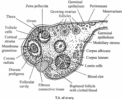
Draw a labelled diagram of a section through ovary.
Answer:
Question 18.
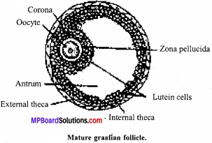
Draw a labelled diagram of a graafian follicle.
Answer:
Question 19.
Write the functions of the following :
- Corpus luteum
- Endometrium
- Acrosome
- Sperm tail
- Fimbriae
Answer:
The functions of the following:
1. Corpus luteum secretes large amount of progesterone which is essential for the maintenance of endometrium of the uterus.
2. Endometrium is necessary for the implantation of the fertillised ovum, for contributing towards making of placenta and other events of pregnancy.
3. Acrosome is filled with enzymes that help in dissolving the outer cover of the ovum and entry of sperm nucleus.
4. Sperm tail facilitates motility of the sperm essential for reaching the ovum to fertilise it.
5. Fimbriae are finger – like projections at the mouth of fallopian tubules that help in collection of the ovum after ovulation.
Human Reproduction Long Answer Type Question
Question 1.
Describe the structure of a seminiferous tubules.
Answer:
Seminiferous tubules are highly coiled tubes, which are lined on the inside by:
1. Male germ cells called spematogonia that undergo meiotic division to form sperm cells.
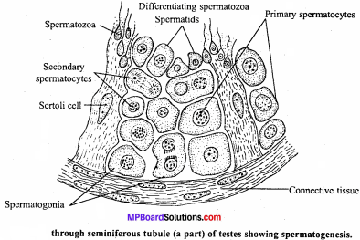
2. Sertoli cells provide nutrition and molecular signals to the germ cells.
Question 2.
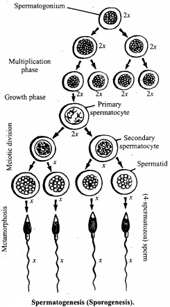
What is spermatogenesis? Briefly describe the process of spermatogenesis.
Answer:
Spermatogenesis:
Formation of sperms in the testis is called as spermatogenesis. It involves in the following steps:
1. Multiplication phase:
In this phase, sperm cells are formed in testes. The inner layer of seminiferous tubules of testes is formed of germinal epithelium. Some of these cells called primary germ cells divide mitotically into spermatogonia which become separated in the germinal layer. Other cells of this layer serve as nutrition for the dividing cells.
2. Growth phase:
In this phase, spermatogonia starts growing, absorbing nutrient substances. These large cells are called primary spermatocytes.
3. Maturation phase:
It is a very important phase. Primary spermatocytes divide twice. The first division is meiotic due to which the number of chromosomes is reduced to half. In this process, primary spermatocyte divides into two halves which are known as secondary spermatocytes. The second division is mitotic and no change takes place in the number of chromosomes. Thus, from two secondary spermatocytes four spermatids are formed. In this manner from one primary spermatocyte four spermatids are formed. These spermatids change into sperm cells of spermatozoa by a process called metamorphosis.
Question 3.
Draw a labelled diagram of human sperm and explain its defferent parts.
Answer:
Structure of sperm:
A mature sperm is a delicate microscopic, motile structure. A typi¬cal mammalian sperm consists of the following three parts:
- Head
- Middle piece and
- Tail.
1. Head:
Head is knob like terminal part of the sperm. It is composed of a large nucleus and an acrosome. At the time of entry of the sperm into egg acrosome secretes spermlysin which dissolve the egg membrane and thus facilitates entry of sperm into the egg or ovum.
2. Middle piece:
It is short and lies between head and tail. It contains two granules called the proximal and distal centrioles in front side and towards posterior side cylindrical middle part of sperm. It is considered as the power house of sperm as it contains compact mass of mitochondria, which provides energy for metabolism and movement of sperm.
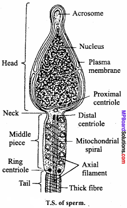
3. Tail:
It is situated on posterior part of sperm. It moves with the help of axial filament. The posterior part of the tail is called as end piece and it is not covered by membrane.
![]()
Question 4.
What are the major functions of male accessory ducts and glands?
Answer:
The main functions of male accessory ducts and glands are as follows:
- Rete testis : They transport sperms from seminiferous tubule to vas efferentia.
- Vas efferentia : Transports sperms to epdidymis.
Question 5.
Describe oogenesis with suitable diagram.
Or
Explain the different phases of oogenesis with ray diagram.
Or
What is oogenesis? Describe its various phases.
Answer:
The process of formation of ova from oogonia in the ovaries is called oogenesis. It consists of three phases:
- Multiplication phase
- Growth phase and
- Maturation phase.
1. Multiplication phase:
The primordial germ cells divide by mitosis to produce oogonia. These oogonia divide by repeated mitotic divisions forming clusters of oogonia called ovigerous cords. These lie close to germinal epithelium. When oogonia stop dividing they are called oocytes. In each cluster of oocytes only one enters to the growth phase and is known as primary oocyte, while the remaining oocytes form follicle cells and provide nourishment to the developing ovum, the primary oocyte.
2. Growth phase:
Growth phase of primary oocyte is very long. It varies from a few days to many years. It takes about 6 – 14 days in hen after ovulation, but three years in frog and in women all the oocytes are present at the time of birth but no one grows till the attainment of puberty, i.e., 12 – 14 years. However afterwards they grow one by one. During growth phase, following changes occur:
(i) Increase in size:
The primary oocyte increases many folds. For example, primary oocyte of the frog in the beginning has a diameter of about 50μ and on maturation it is about 1000μ to 2000μ. In man also on maturation oocyte increases in size about 7 times. The size increases due to the accumulation of reserve food like proteins and fats in the form of yolk. Due to heavy weight it is usually concentrated towards and lower portion of the egg forming vegetative pole. The portion of the cytoplasm with egg pronucleus remains often separated from the yolk and occurs towards the upper portion of the egg forming animal pole.
(ii) Number of mitochondria increases and in certain cases (birds, amphibia) they are concentrated in the same places to form mitochondrial clouds.
(iii) Increase the amount of ER and activity of golgi complex.
(iv) Synthesis of yolk or Vitellogenesis:
Chemically, yolk is a lipoprotein composed of protein, phospholipids and neutral fats along with little quantity of glycogen. The yolk is synthesized in the liver of female in soluble form and is transported through circulation to the follicle cells surrounding maturing ovum. From follicle cells it is absorbed by the ovum and is deposited in the form of yolk granules and platelets in the ooplasm. The mitochondria and golgi complex are said to be responsible for the conversion of soluble yolk into insoluble granules or platelets.
(v) Formation of thin vitelline membrane around the oocyte.
(vi) Increase the size of nucleus:
Due to the increase in the amount of DNA, nuclear sap and number of nucleoli, nucleus increases in size.
(vii) Gene amplification:
In growth phase the nucleolar genes which code for ribosomal RNA and are located in the nucleolar organizer region multiply to facilitate rapid synthesis of ribosomal RNA. This multiplication of genes without mitosis is known as gene amplification or redundancy.
3. Maturation phase:
In this phase, the nucleus of oocyte undergoes two maturation divisions. The first division is meiotic, as a result two haploid (n) cells are produced. In this division, cytokinesis is unequal, the large daughter cell with almost entire cytoplasm and yolk forms the secondary oocyte. While the smaller one with haploid nucleus (n) and almost without cytoplasm forms the first polar body which is given off from the surface of oocyte at the animal pole. The secondary oocyte with haploid number of chromosomes undergoes second maturation division or second meiotic division (it is meiotic division).
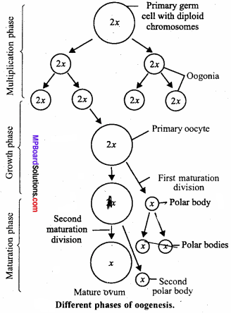
This division is also unequal the large one containing yolk is called ovum and small cell is the second polar body. The first polar body may also divide thus, producing total three bodies which degenerate soon. So, as a result of oogenesis only one functional ovum is formed from a primary oocyte. In most of the vertebrates, the first meiotic division occurs with the commencement of the growth phase, and second maturation division occurs when egg is activated by the entry of sperm.
Question 6.
Describe the structure of ovary.
Answer:
These are a pair of female gonads or primary sex organs lying one on each side of uterus. Ovary is attached to abdominal wall as well as utems by means of ligaments. It is surrounded by a fold of peritoneum named mesovarium. Ovary is internally differentiated into four parts : Germinal epithelium, tunica albuginea, cortex and medulla.
Germinal epithelium is the outermost layer of cuboidal to flattened cells. Germinal epithelium is followed by a sheath of condensed connective tissues which is termed as tunica albuginea. It is followed b.y cortex. The central part of ovary contains medulla. A large number of groups of specialized cells are present in the cortex which are termed as ovarian follicles. These follicles are found in four developmental stages.
1. Incipient follicle:
The central part of these follicles contains a large cell which is covered by many smaller cells.
2. Primary follicle:
These follicles are developed from incipient follicles.
3. Vascular follicle:
It is formed from primary follicles. The oocyte of these follicle is covered by a many layered thick wall.
4. Graafian follicle:
Mature ovarian follicle is termed as graafian follicle. It is covered by two sheaths derived from cortex. The follicle contains a single oocyte. A group of follicular cells surrounds the oocyte or ovum. It is called cumulus ovaricus. Another group produces membrane granulosa. Oocyte has two noncellular membranes, inner vitelline membrane and outer zona pellucida.
Note:
Question 7.
Describe the process of fertilization and give its significance.
Answer:
Fertilization:
Fertilization is the fusion of male and female gametes to produce a single diploid cell, called zygote. Fertilization in human female takes place in fallopian tube. In a sexual mating or coitus the male ejaculates semen into the vaginal passage of the female using the copulatory organ, the penis. In a single coitus as many as 200 million sperms are introduced into the female genital tract but out of this huge number of sperms only one is destined to fuse with the ovum, provided the fallopian tube lodges of fully developed secondary oocyte.
Sperms travel through the vaginal passage and enter the uterus through the cervix. They travel further through the uterus and finally enter the fallopian tube.The vaginal passage is highly acidic to prevent any bacterial infection but this acidic medium is not suitable for the survival of sperms. Many sperms die on the way. The liquid medium of the semen is alkaline which can neutralize the acidity of vagina to some extend and keep the sperms alive and active. The sperms move with the speed of 1-5 to 3 mm per minute.
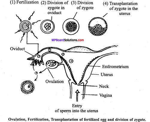
The ovum gets surrounded by a large number of sperms but usually only one fuses with the ovum. The sperm penetrates the ovum using certain chemical substances of enzymatic nature.These chemicals are called spermlysins which are present in the acrosome. Certain receptors on the cell surface of the ovum enable the sperms to penetrate the wall of ovum. The ovum is surrounded by follicle cells.
These cells are joined together by a glue like substance known as mucopolysaccharide, an acid, called hyaluronic acid. The sperm produces spermlysin, known as hyaluronidase. The over all changes in a sperm before the fertilization is called capacitation. The hyaluronidase enzyme facilitates the sperm to pen-etrate through the corona radiata (follicle cells), zona pellucida and the plasma membrane of ovum. The nucleus and the cytoplasmic components get inside the ovum, leaving the tail outside. The entry of sperm stimulates the ovum and the signal is transmitted to the egg surface incapacitating all the other cells surrounding the ovum.
Nucleus of sperm move towards the nucleus of ovum and they fuses with each other to form zygote. It takes 2 – 2\(\frac { 1 }{ 2 }\) hours to complete fertilization process. Once the ovum has been fertilized a barrier forms around it that normally prevents other sperms from entering. Now fertilized egg reaches to the uterus and within seven days of fertilization it is transplanted to the wall of the uterus.
Significance of Fertilization:
- Egg becomes active after entry of sperm and completes its second maturation division.
- Formation of fertilization membrane prevent entry of other sperms in the ovum. In human this membrane is not formed.
- It restores the diploid number (2n) of chromosomes in the zygote.
- It combines characters of male and female resulting in the introduction of varia-tions. These variations make the offspring better equipped for struggle against environmental conditions to ensure the existence.
- The ovum is stimulated to cleavage.
- After fertilization ovum rotate inside the membrane.
- Ovum do not contain centriole and obtain it from sperm during this process thus zygote continuously divides.
- It is necessary for the egg to attain maturity.
![]()
Question 8.
Describe development of embryo up to the formation of three germ layer. Give the names of organs formed by three germ layer.
Or
Define cleavage. Describe the changes that occur in embryo till gastrulation.
Answer:
The term cleavage refers to a series of rapid mitotic divisions of the zygote following fertilization forming a many celled blastula. Following are the various steps of embryonic development in human up to the formation of three germ layer:
1. Formation of morula:
The fertilized zygote divides. It undergoes successive quick mitotic cell divisions called cleavage. First cleavage is holoblastic, unequal and meridional. It divides the oyum completely into two unequal blastomeres. The plane of cleavage passes
through animal vegetal axis, i.e., it is meridional. Large blastomere divides little earlier than the small one giving a transitional three cell stage.
As a result of further cleavages, a solid mass of mulberry shaped embryo is formed called morula. Morula is of the size of zygote but consists of 32 cells. These cells are of two types , The outer layer of smaller cells around, inner larger cells. Within 72 hours of fertilization, morula reaches the uterus.
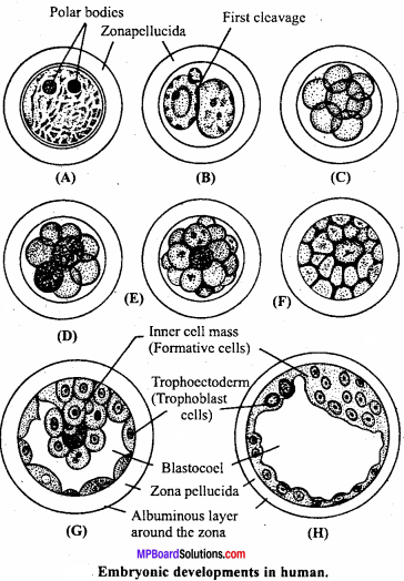
2. Formation of blastula:
Transformation of morula into a blastula starts by the rearrangement of blastomeres. This leads to the formation of a central cavity inside the morula. The outer cells of morula absorb the nutritive fluid secreted by the uterine mu¬cous membrane and transform into trophoblast. The fluid absorbed by these cells collects in the central cavity called biastocoel. This cavity separates the trophoblast from the inner mass of larger cells except on one side and is termed blastocyst. The inner cell mass is pushed to one pole as a small knob. This knob gives rise to the embryo and is termed as embryonal knob.
3. Formation of gastrula:
In this embryonic stage development of three germ layer occurs. In this stage morphogenic movement of cells of embryo occurs, as a result of this three germ layers are formed. A cavity develops at the centre called as archenteron which opens outside through blastopore.
4. Formation of three germ layers:
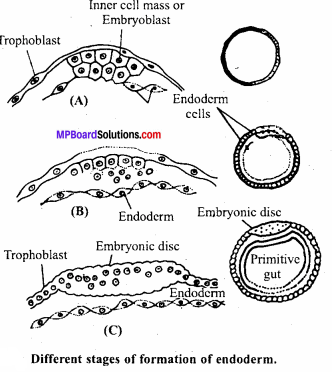
Formation of endoderm:
The enlargement of blastodermic vesicle is followed by the separation of few cells from the inner cell mass. These cells push out from the blastocoel to become the initial cells of the innermost layer of gastrula, the pattern of a tube within a tube. The inner tube is called primitive gut. It is differentiated into gut tract which is within the body and a yolk sac that communicates with the gut of the embryo. The remaining cells of the inner cell mass are organized to form the embryonic disc.
Formation of mesoderm:
After the formation of endoderm an increased rate of cell proliferation takes place at the caudal end of the embryonic disc. This results in the localised increase in the thickness of the disc. These cells subsequently get detached from the embryonic disc and get organized to a well demarcated mesodermal layer.
Formation of ectoderm:
After the formation of endoderm and mesoderm, the remaining cells of the embryonic disc arrange themselves in a layer to form ectoderm.
![]()
Question 9.
Write differences between spermatogenesis and oogenesis.
Answer:
Differences between Spermatogenesis and Oogenesis:
Spermatogenesis:
- Sperms are produced by this process.
- In this process primary spermatocytes are formed by maturation of germinal epithelium cells.
- Primary spermatocyte divides to form four spermatids.
- There is equal division.
- Large number of sperms are formed by this process.
Oogenesis:
- Ovums are produced by this process.
- In this process primary oocytes are formed by maturation of germinal epithelium.
- Primary oocytes divides to form one ovum and three polar bodies.
- There is unequal division.
- Less number of ovums are formed by this process.