Students get through the MP Board Class 11th Biology Important Questions Chapter 21 Neural Control and Coordination which are most likely to be asked in the exam.
MP Board Class 11th Biology Important Questions Chapter 21 Neural Control and Coordination
Neural Control and Coordination Class 11 Important Questions Very Short Answer Type
Question 1.
Write the name, nature, origin and distribution area of that nerve which rotates tongue.
Answer:
Hypoglossal nerve rotates the tongue. It is stimulative in nature and originates from the medulla of brain and goes up to the muscles of tongue.
Question 2.
What would be happened in the absence of acetylcholine from synapse?
Answer:
In synapse, acetylcholine transmits the nerve impulse from the axon of one nerve cell to the dendrite of the other nerve cell. Thus in the absence of acetylcholine from synapse nerve impulses do not pass from one nerve cell to other and thus transmission of nerve impulses is inhibited.
Question 3.
Give one example of reflex action.
Answer:
Withdrawal of hand when it is pricked with a needle.
Question 4.
What are axons?
Answer:
The largest branch (dendrite) of nerve cells is known as axon. It plays an important role in the conduction of informations.
Question 5.
Name the humours found in the eyes and describe their functions?
Answer:
Eye has two types of humours :
- Aqueous humour: It is a fluid which is filled in aqueous chamber present between the cornea and lens. It
protects the lens, iris and conjunctiva from direct light and outer forces. - Vitreous humour: It is jelly like liquid which is filled in vitreous chamber present between lens and retina. It provides an specific shape to eye ball and also protects it from adherence.
Question 6.
Describe the functions of the following :
(i) Cerebellum,
(ii) Iris,
(iii) Eustachian tube.
Answer:
(i) Cerebellum: The part of the brain which lies behind and below the cerebrum.
(a) Maintenance of posture and equilbrium of the body.
(b) Maintenance of muscle tone.
(c) It modulates and moderates voluntary movements initiated by cerebellum.
(a) It is the site of pain, touch and temperature.
- It controls voluntary movement of muscles, visual and audio sensation.
- Iris: It controls the amount of light that enters the eyes.
- Eustachian tube: It controls the pressure applied on tympanum (membrane between external and middle ears).
![]()
Question 7.
What is synapse?
Answer:
Nerve ending of axon of one neuron are connected with the dendrites of next neuron by specific joint is called as Synapse.
Question 8.
Name five sense organs and give their functions.
Answer:
| Sense organs | Functions |
| (a) Skin | Touch |
| (b) Eye | Vision |
| (c) Ear | Hearing and balance of the body |
| (d) Nose | Smell |
| (e) Tongue | Taste |
Question 9.
Give nature of following nerves :
(a) Olfactory,
(b) Vagus,
(c) Auditory,
(d) Mandibular.
Answer:
| Nerve | Nature |
| (a) Olfactory | Sensory |
| (b) Vagus | Motor and sensory |
| (c) Auditory | Sensory |
| (d)Mandibular | Motor. |
Question 10.
Why owl cannot see during day time but it can see during night ?
Answer:
Owl cannot see during day time as cones are absent in their retina. But it can be able to see during night because their retina possessing a large number of rods.
Question 11.
(a) Which part of the ear determine pitch of the sound ?
(b) Which is the most developed part of the human brain ?
(c) Which part of the central nervous system functions as master clock ?
Answer:
(a) Internal ear,
(b) Cerebrum,
(c) Brain.
Neural Control and Coordination Class 11 Important Questions Short Answer Type
Question 1.
Give functions of cerebrum of man.
Answer:
Functions of cerebrum :
- Its parietal lobe is the centre of pain, touch and stimuli of temperature.
- Its motor area of frontal lobe regulates movement of voluntary muscles.
- Its premotor area of frontal lobe regulates all the functions regulated by autonomous nervous system.
- Its temporal lobe feels taste and smell.
- Visual area of occipital lobe is responsible for the knowledge of light and dark.
- Hypothalamus regulates hunger, thurst and quantity of urine. It also regulates secretion of anterior lobe of pituitary gland.
Question 2.
Write the names of brain ductules of human beings.
Answer:
Human brain consists of a hollow cavity from which the cavity of forebrain is called as lateral cavity. It represents first and second cavity. The cavity of diencephalon is called as third or tertiary cavity. The cavity of mid-brain is called as oieter whereas the cavity of hind-brain is known to be as fourth cavity.
Question 3.
Name the smallest bone of human ear. What happen if this bone is removed ?
Answer:
The smallest bone of human ear is stapes. This bone transmits sound vibration from the tympanic membrane to the membrane of the oval window. This bone increases the force of vibration thus acting like lever, when stapes is removed from ear the waves of sound do not enter the inner ear because stapes which is associated with fenestra ovalis, transmits the vibration of sound into perilymph of vestibular cavity.
Question 4.
What are adrenalin and acetylcholine? Give their functions.
Answer:
Adrenalin and acetylcholine are chemical substances secreted by sympathetic and parasympathetic nerve fibre. Both the chemicals function in opposite way. Adrenalin induces various activities and acetylcholine inhibits them.
Question 5.
What is mosaic vision?
Answer:
This type of vision is found in cockroach because they have compound eyes. Many small images are formed by them. Complete vision is compound form of these many small images and is called as mosaic vision.
Question 6.
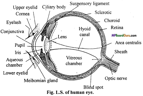
Draw a well labelled diagram of longitudinal section of human eye.
Answer:
Question 7.
What is hypermetropia and myopia? (M.P. 1995 Set II)
Answer:
Hypermetropia (Far or Long-sightedness): It is a disease of eyes in which light rays strike the retina before being converged to form image i. e. image is formed beyond the retina. This happens because of (i) abnormally small eye ball, (ii) flattening or abnormally low convexity of the lens. Hypermetropia is corrected by using spectacles with suitable biconvex or convergent lenses.
Myopia (Short sightedness) : This defect results from abnormally long eye ball or abnormally high convexity of lens. The eyes are able to observe nearby objects clearly. However distant objects like writing on the black board or a bird sitting on a tree are not visible properly. It is because of the light rays from distant objects do not form the images on the retina but in front of it in the vitreous humour. The defect is corrected by using spectacles having suitable concave or divergent lens.
![]()
Question 8.
What do you understand by nervous system? Nervous system is divided into how many parts?
Answer:
The system which recieves the stimulus, transmits it to the other part of the body and the corresponding effects are shown is known as nervous system.
Nervous system consist of the following three parts :
- Central nervous system : Comprising of brain and spinal cord.
- Peripheral nervous system : Includes the nerves which arise from CNS.
- Autoinomic nervous system : Has connections with central nervous system, but works somewhat independly to regulate the involuntary activites of the body e.g. Heart beat, peristalsis etc.
Question 9.
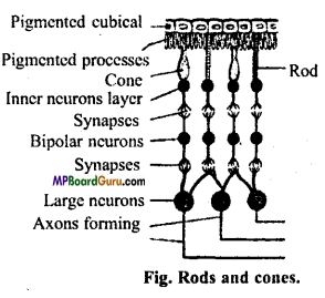
Explain the role of rods, cones and yellow spot in human eye. (MP 2000, 02, 06 Set C,)
Answer:
1. Rods : Rods are dark light receptors containing rhodopsin synthesized from vitamin A.
Rods are responsible for photopic vision and function best at night and dim light.
2. Cones: Cones are colour receptors containing iodopsin. They function in strong light and responsible for scotopic vision (colour vision).
3.Yellow spot: Posterior part of retina contains only cones and rods are absent and contains yellow pigment called yellow spot. It helps in the formation of clear picture of object.
Question 10.
How many types of nerves are there on the basis of their functions?
Answer:
According to the functions neurons are divided into three parts :
- Sensory Nerves: These nerve fibers conduct impulses from receptor organ (such as nervous) towards the central nervous system.
- Motor nerves : These nerve fibres carry the impulses from CNS to effector organs.
- Mixed nerves: These nerves contain both sensory and motor nerve fibres, hence they do both the functions.
Question 11.
Differentiate the following : (NCERT)
(a) Central Nervous system and peripheral Nervous system.
(b) Resting Potential and Active potential.
(c) Choroid and Retina.
Answer:
(a) Central Nervous System (CNS): Originates from ectoderm part of the embryo. It consists of Brain and spinal cord. Neural tube part of the embryo develops to form this system. This part is responsible for processing of information, where as Peripheral Nervous system (PNS) includes the nerves which arise from the central nervous system.
Nerves arises from brain are called as Cranial nerves and nerves arises from spinal cord are called as spinal nerves. It fonns a communicating system by connecting central nervous system with sensory organs (receptors) through sensory nerves and effector organs through motor nerves.
(b) Resting Potential and Action Potential: At resting stage difference between electric potential of plasma membrane is called as Resting potential. Its value is -70mv, whereas whenever any part of the axon is stimulated depolarisation of Axolemma occurs, thus potential difference decreases up to 30 mV. This is called as Action potential.
(c) Choroid and Retina : Choroid is the middle or second coat of the wall of the eye made up of connective tissue, which is highly vascularised and pigmented. In the front side it fonns iris which has a hole in the centre called as pupil, where as Retina is the innennost layer of the eye wall, which consists of light sensitive cells called as rods (which perceives light) and cones (which distinguish colours).
![]()
Question 12.
Explain the structure and functions of spinal cord with diagram.
Or,
Draw a well labelled diagram of T. S. of spinal cord.
Answer:
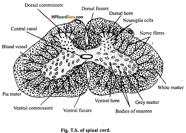
Structure of spinal cord: The spinal cord consists of central canal surrounded by a central core of grey matter, which is surrounded by white matter. In transverse section, grey matter shows a butterfly shape, i. e., it is produced into two dorsal horns and two ventral horns. A central canal is present in the centre of the grey matter. The spinal cord serves as a centre for spinal reflexes and as pathways to and’from the brain.
The grey matter contains the bodies of neurons with tree-like branching of their dendrons and neurological cells. The white matter is composed of obliquely running medullated nerve fibers supported by pro¬longations of the neuroglia. The bands of fibers which extend transversely, one dorsal and other ventral, to the central canal are known as dorsal and ventral commissures, respectively, Thirty one pairs of spinal nerves arise from the spinal cord. Each nerve is attached to the spinal cord by dorsal and ventral root.
The sensory nerve fibres enter the spinal cord by way of dorsal root while the motor fibres emerge from the spinal cord by way of the ventral root to conduct motor impulses through spinal nerves to effector organs (gland and muscles).
Functions of spinal cord :
(i) Spinal cord controls and coordinates the reflex actions.
(ii) It acts as link between spinal nerves and brain and conducts the impulses of brain.
Question 13.
Write differences between :
(a) Yellow spot and Blindspot.
(b) Short sight and Long sight.
(c) Endolymph and Perilymph.
Answer:
(a) Differences between Yellow spot and Blind spot
| Yellow spot | Blind spot |
| 1. Posterior part of retina has yellow pigment. There is a point in retina where the It is called yellow spot. | optic nerve fibre leave the retina. This point is called blind spot |
| 2. In yellow spot, rod and cones are present. | In blind spot, rod and cones are absent. |
(b) Differences between Short sight and Long sight
| Short sight | Long sight |
| 1. In this type nearby objects are clearly visible, but the distant objects are not visible but clearly visible. | In this type, distant objects are clearly nearby objects are not clearly visible. |
| 2. Image is formed infront of retina. | Image is formed behind the retina. |
| 3. This defect is removed by using concave lens. | This defect is removed by using convex lens. |
(c) Differences between Endolymph and Perilymph
| Endolymph | Perilymph |
| 1.The membranous labyrinth and cochlea are filled with a fluid called as endolymph which contain CaC03 particles called as otoliths, | Space between bony labyrinth and membranous labyrinth is filled with a fluid called as perilymph, which is a cerebrospinal fluid. It protects internal ear. |
| Sound wave causes vibration of endolymph, due to which otoliths vibrate. Vibration of endolymph helps to receive stimuli of sound, thus helps for hearing as well as balancing. |
Neural Control and Coordination Class 11 Important Questions Long Answer Type
Question 1.
Describe following processes : (NCERT)
(a) Polarisation of membrane of nerve fibre.
(b) Depolarisation of membrane of nerve fibre.
(c) Conduction of impulse through nerve fibre.
(d) Transmission of impulse across synapse (chemically).
Or,
What is nerve impulse? How is it conducted?
Answer:
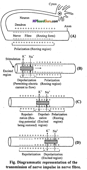
Nerve impulse: The sum total of the physical and chemical reactions which take place in the propagation of the wave of physiological activity along the nerve fibre is called as nerve impulse.
Propagation of Nerve impulse :
1. Depolarization: When a stimulus of any kind electrical, mechanical or chemical acts on the nerve fibre momentarily, a local increase occurs in the permeability of membrane by opening sodium gate at the site of stimulus. This suddenly brings sodium ions influx into the cell, carrying enough positive charges to the inside to cause complete disappearance of the normal resting potential and usually enough charges to develop a positive state inside the fibre. This positive state of the fibre is called reverse potential. It results in the depolarization of the membrane.
2. Action potential: The changed electrical potential of the plasma membrane of neuron is called action potential. The initial change produces an ionic imbalance across the membrane on either side of the points of stimulus producing local electrical current. These areas of negative depolarization in turn initiate changes in the membrane adjacent to them. A wave of electric charges or depolarization along the length of the nerve fibre or axon is called nerve impulse.
3. Repolarization: With the increase of positive charge inside further entry of Na+ is prevented and the permeability of membrane decreases. Na+ ions are pushed out which transfer large number of positive charge outside, once again creating negativity inside the membrane which restores the original resting potential. This is known as repolarization. The repolarization starts exactly on the same spot where depolarization had started and then continues to advance ahead. The entire process of repolarization requires some time during which the nerve cannot be stimulated again. It is known as refractory period.
Theories of nerve impulse conduction or Transmission :
1. Saltatory conduction or through nerve fibre : Myelin sheath acts as insulator in the medullated nerve fibres. It is almost impermeable to the ions. The membrane at the nodes of Ranvier is 500 times more permeable. The nerve impulse in these fibres passes from node to node along the entire length of nerve fibre rather than continuously along the entire fibre as occurs in unmyelinated fibre.
This process is called saltatory conduction. It is of dual value. First by causing the depolarization process to jump along nodes of Ranvier on the axis of the nerve fibre increases greatly the speed of impulse travel and secondly saltatory conduction conserves energy for the axon because only the nodes depolarize.
The velocity of conduction in nerve fibre varies from 5 metres per second in a small unmyelinated fibres to as high as 130 metres per second in large myelinated fibres.
Transmission of impulse across synapse (synaptic transmission): The area where the dendrite of one neuron is in close proximity with the axon of another is known as the synapse. The space between the axon and dendrite is called the synaptic cleft.
When the impulse reaches the tip of axon it stimulates the synaptic vehicles which move towards the synaptic cleft and discharge the neurotransmitter or neurohormones.
They then return the knob and become recharged. This chemical neurotransmitter substance has been identified as acetylcholine. Once released the neurotransmitter diffuses across the synaptic cleft and becomes attached to chemo- receptors present on the dendrites of the next neuron.
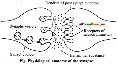
This attachment triggers the depolarization of the membrane and initiates action potential which travels across the next neuron. Thus, the actual transmission of the impulse across the synapse is by means of specific chemicals. Just after the transmission of impulse through the synapse, acetylcholine is hydrolyzed into acetic-acid and choline by the enzyme acetylcholinesterase present in the post synaptic membrane. The membrane again becomes polarized, so another impulse must come down the axon and release more acetylcholine for the transmission of another impulse.
![]()
Question 2.
Give three-three functions of sympathetic and parasympathetic nervous system.
Answer:
Functions of Sympathetic Nervous System :
- Sympathetic nervous system maintains the physical balance during distress conditions.
- It stimulates the adrenal glands to secrete adrenalin. Thus, sympathetic nerves function with the adrenal glands as a well-integrated sympathetic-adrenal system having widespread effects.
- This system is responsible for the increase in the rate of heartbeat and blood pressure.
- It reduces the peristaltic movement of alimentary canal.
- It brings about the extension of pupil.
- It contracts the sphincter muscles of urinary bladder.
- It increases the sugar level in blood.
- It increases the number of RBCs in blood.
Functions of Parasympathetic Nervous System :
- Parasympathetic nervous system opposes the effects of the sympathetic autonomic system.
- It decreases rate of heartbeat.
- It decreases blood pressure.
- The nerve ending of this system secrete acetylcholine.
- It constricts pupil of the eyes.
- It relaxes the muscles of root hairs.
- It contracts the muscles of ureter.
- It increases the secretion of saliva and digestive juices.
Question 3.
Name the regulating centre of following in brain :
(a) Temperature,
(b) Hunger,
(c) Respiration,
(d) Water balance,
(e) Vision,
(f) Hearing,
(g) Menstrual cycle,
(h) Learning, (i) Pituitary gland,
(j) Coordination and equilibrium,
(k) Transport,
(l) Sleep,
(m) Heartbeat.
Answer:
(a) Temperature – Thalamus
(b) Hunger – Hypothalamus
(c) Respiration – Medulla oblongata
(d) Water balance – Hypothalamus
(e) Vision – Cerebral cortex
(f) Hearing – Cerebral cortex
(g) Menstrual cycle – Hypothalamus
(h) Learning – Cerebral cortex
(i) Pituitary gland – Hypothalamus
(j) Equilibrium and Coordination – Cerebellum
(k) Transport – Medulla oblongata
(l) Sleep – Cerebral cortex
(m) Heartbeat – Medulla oblongata.
Question 4.
Describe following structures in breif. (NCERT)
(A) Brain,
(B) Eye,
(C) Ear.
Answer:
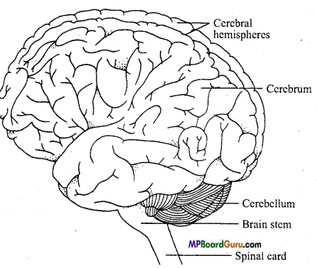
(A) Brain: Mammalian brain is divisible into three parts :
1. Fore-brain or prosencephalon consists of cerebrum (telencephalon), olfactory lobes (rhinencephalon) and diencephalon (thalamencephalon).
2. Mid-brain or mesencephalon consists of optic lobes (corpora quadrigemina) and cerebral peduncle.
3. Hind-brain or rhombencephalon formed of cerebellum (metencephalon), medulla oblongata (myelencephalon) and pons varoli.
(1) Forebrain :
(i) Olfactory lobe : In mammals the olfactory lobes are located at the anterior end of brain, ventral in position. They are reduced and covered by cerebral hemisphere. Each lobe is further differentiated into olfactory bulb and olfactory tract. The olfactory tract ends in a rounded elevation, the tuberculum olfactory. But in human brain due to the over-development of cerebrum the olfactory lobes get incorporated with the cerebrum and are visible only in the ventral view. Olfactory lobes are concerned with the sense of smell. The nature of the nerve arising from the olfactory lobes are purely sensory.
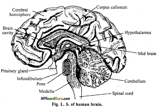
(ii) Cerebrum: Cerebrum is the largest part of the brain. It is made up of two halves, the right and the left cerebral hemispheres. A deep median longitudinal groove is the demarkation between two halves. The two halves are connected by means of a broad transverse band of white matter called corpus callosum or cerebri.
Deep furrows divide each cerebral hemisphere into frontal lobe, temporal lobe, occipital lobe and a small lobe called insula. Insula is a triangular eminence lying deeply in lateral fissure of temporal lobe. The other lobes are named according to the position they occupy. The surface of cerebrum is folded, having elevation and depressions called sulci and gyri that increases surface area.
(iii) Diencephalon: It is a small area, it remains covered with cerebrum. It consists of following:
(a) Epithalamus: It forms the roof of diencephalon having anterior choroid plexus concerned with supply of nutrition of brain and vestigial pineal body.
(b) Optic thalami: These forms lateral walls of diencephalon and contain optic fibres which go towards cerebrum.
(iv) Hypothalamus : The base of the brain constitutes the hypothalamus. It forms the floor of diencephalon. It has a hypophysis or master gland or pituitary gland, which is an endocrine gland. Anterior to it is a geniculate body which relay optic impulses to cerebrum. An optic chiasma is formed by the crossing of optic fibres. The hypothalamus, consists of large amount of grey matter present within the white matter. It contains the nerve centres for control of temperature, hunger, thirst and emotions, sleep and sexual activities. It also produces various neurohormones that control the secretion of anterior pituitary. Hormones of posterior pituitary are produced in hypothalamus which reach the pituitary by hypophyseal portal vessels.
2. Mid-brain: The mid-brain or mesencephalon joins the cerebrum with the cerebellum. It consists of four lobes called quadrigemina. Anterior pair of lobes is concerned with vision and posterior ones with hearing. It has large number of nerve cells scattered within the white matter. These nerve cells are involved in controlling the voluntary muscle activities and also act as centres for auditory and visual reflexes. It can modify some motor activities initiated by cortex.
3. Hind-brain: The hind brain or rhombencephalon consists of cerebellum, pons and medulla and many visceral functions forms its major part and is a large reflex centre for the co-ordination of activities of voluntary muscles.
The brain-stem consists of pons varoli in front of cerebellum and behind it medulla oblongata is present. Pons varoli runs transversely and carries impulses from one portion of cerebellum to another. The medulla oblongata lies in between the pons varoli and spinal cord and controls various reflexes like breathing, swallowing, salivation, chewing, coughing, sneezing etc.
(B) Human Eye: L.S. of human eye : Human eye is a spherical ball like structure filled with a vitreous liquid. It is covered by upper and lower eyelids. The eyeball of human beings is made up of from the following three layers :
1. Sclerotic layer: It is the outermost layer of eye. Their 1/3 transparent part is called as cornea. Covering of cornea is a thin transparent membrane called conjunctiva. Cornea permits light to enter the eye.
2. Choroid layer: It is an extensive plexus of small blood vessels embedded in loose connective tissue. At the anterior margin of the choroid a greatly thickened structure projects immediately into the eyeball towards the margin of the lens.
This portion of the choroid is called as ciliary body. Anterior to the ciliary body the vascular tunic is thin and constitute the iris of the eye; in the middle of the iris is a round opening called the pupil. A lens (biconcave) is present below the iris which is encased in a strong fibrous lens capsule and containing a high concentration of protein.
3. Retina: It is the innermost part of the eye which lines the inside surface of the posterior two third of the eyeball and lies on the inner surface of the choroid. The retina containing light sensitive cells called rods and cones that detect light and colour and then send appropriate signals to the brain to cause vision.
Two functions of rods and cones are :
- The photoreceptor, rod cells of retina contain rhodopsin. They function in dim light and enable us to see in darkness at night.
- The photoreceptor, cone cells of retina contain iodopsin. They function in day light and artificial bright light. They are associated with perception of colours
Note: For

(C) Ear: Human ear is highly developed. It can be divided into three parts :
1. External ear: It consists of flap like structure called pinna which collects sound and pass it to the auditory canal, at the end of which tympanic membrane is found.
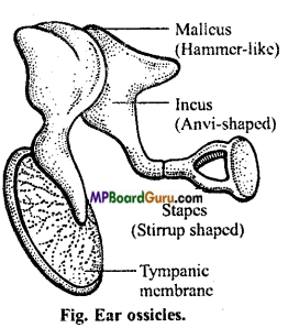
2. Middle ear: It consists of tympanic cavity which is present between the tympanic membrane and the internal ear. Three small bones called ear ossicles are found in the middle ear. The first bone, which is club-shaped and is attached at one end to the tympanic membrane is called malleus.
Its other end is attached to an anvil-shaped bone called incus. The narrow end of incus is attached to the bone called stapes which is stirrup-shaped and has an aperture in it.
The other end of stapes is fixed to the membrane on the fenestra ovalis. In mammals, all these three bones originate from the bones of the jaws, malleus from articular, incus from quadrate and stapes from hyomandibular. The tympanic cavity is connected with the pharynx by means of eustachian tube.
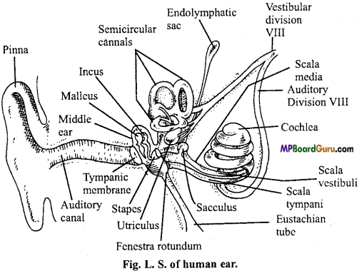
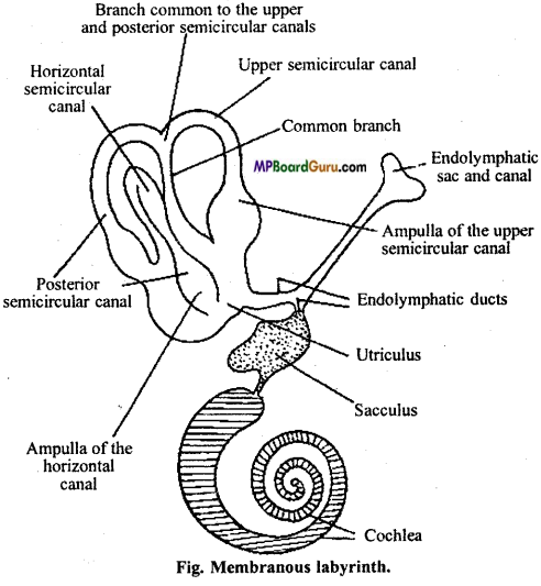
3. Internal ear: The membranou s labyrinth of internal ear consists of two chambers : utriculus and sacculus. These chambers are connected with each other by a narrow canal called saccularicular canal. Three semicircular canals arise from the utriculus. According to their position, they are known as anterior, posterior and external or horizontal semicircular canals. The anterior and posterior semicircular canals arise together and run for a short distance and then separate. After taking a curved course, their other ends again open into the utriculus where each of them enlarges to form an ampulla. Each ampulla contains a sensory crista which is formed of ciliated and supporting cells.
Two such cristae maculae are present in sacculus and one in the utriculus. The hind part of sacculus is coiled like a spring and is known as cochlear duct. Utriculus, ampullae, semicircular canals, sacculus and cochlear duct are all filled with a thick fluid called endolymph which contains minute particles of calcium carbonate known
as otoliths.
The internal ear is situated in the temporal region of the skull within the periotic bones which take the shape of the internal ear. Thus the whole internal ear, i.e., membranous labyrinth is enclosed in a bony labyrinth.
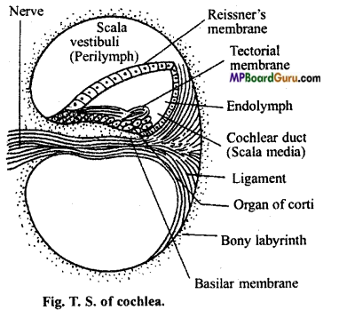
The space between these two is filled with a fluid called perilymph. The cochlea is associated with hearing. It is a bony tube coiled like a conch shell. It is wound around a bony cone of bone, known as central pillar or modiolus. The cochlear nerve passes through the modiolus. The scala media remains separated from the vestibular canal of the bony labyrinth by a vestibular membrane or resisters membrane. It is also separated from the underlying tympanic canal by a basilar membrane.
The basilar membrane possesses several rows of hair cells serving as auditory receptors all along its coiled course. This structure is known as Organ of Corti which is the organ of hearing. It is composed of several specialized cells. Corti’s rods are double row of arching rods based on basilar membrane and ‘forming the spiral tunnel of Corti. Hair cells are the sensory organ of Corti’s organ. Dieters cells are the supporting cells between rows of outer hair cells in organ of Corti. A tectorial membrane lies in contact with the hair cells.
Question 5.
Draw labelled diagrams of following :
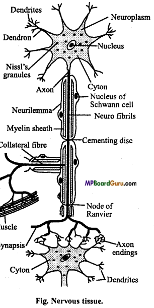
(a) Neuron,
(b) Brain,
(c) Eye,
(d) Ear. (NCERT)
Answer:
(a) Neuron :
(b) Brain:
(A) Brain: Mammalian brain is divisible into three parts :
1. Fore-brain or prosencephalon consists of cerebrum (telencephalon), olfactory lobes (rhinencephalon) and diencephalon (thalamencephalon).
2. Mid-brain or mesencephalon consists of optic lobes (corpora quadrigemina) and cerebral peduncle.
3. Hind-brain or rhombencephalon formed of cerebellum (metencephalon), medulla oblongata (myelencephalon) and pons varoli.
(1) Forebrain :
(i) Olfactory lobe : In mammals the olfactory lobes are located at the anterior end of brain, ventral in position. They are reduced and covered by cerebral hemisphere. Each lobe is further differentiated into olfactory bulb and olfactory tract. The olfactory tract ends in a rounded elevation, the tuberculum olfactorium. But in human brain due to the over development of cerebrum the olfactory lobes get incorporated with the cerebrum and are visible only in the ventral view. Olfactory lobes are concerned with the sense of smell. The nature of the nerve arising from the olfactory lobes are purely sensory.
(ii) Cerebrum: Cerebrum is the largest part of the brain. It is made up of two halves, the right and the left cerebral hemispheres. A deep median longitudinal groove is the demarkation between two halves. The two halves are connected by means of a broad transverse band of white matter called corpus callosum or cerebri.
Deep furrows divide each cerebral hemisphere into frontal lobe, temporal lobe, occipital lobe and a small lobe called insula. Insula is a triangular eminence lying deeply in lateral fissure of temporal lobe. The other lobes are named according to the position they occupy. The surface of cerebrum is folded, having elevation and depressions called sulci and gyri that increases surface area.
(iii) Diencephalon: It is a small area, it remains covered with cerebrum. It consists of following:
(a) Epithalamus: It forms the roof of diencephalon having anterior choroid plexus concerned with supply of nutrition of brain and vestigial pineal body.
(b) Optic thalami: These forms lateral walls of diencephalon and contain optic fibres which go towards cerebrum.
(iv) Hypothalamus : The base of the brain constitutes the hypothalamus. It forms the floor of diencephalon. It has a hypophysis or master gland or pituitary gland, which is an endocrine gland. Anterior to it is a geniculate body which relay optic impulses to cerebrum.
An optic chiasma is formed by the crossing of optic fibres. The hypothalamus, consists of large amount of grey matter present within the white matter. It contains the nerve centres for control of temperature, hunger, thirst and emotions, sleep and sexual activities. It also produces various neurohormones that control the secretion of anterior pituitary. Hormones of posterior pituitary are produced in hypothalamus which reach the pituitary by hypophyseal portal vessels.
2. Mid-brain: The mid-brain or mesencephalon joins the cerebrum with the cerebellum. It consists of four lobes called quadrigeminal. Anterior pair of lobes is concerned with vision and posterior ones with hearing. It has large number of nerve cells scattered within the white matter. These nerve cells are involved in controlling the voluntary muscle activities and also act as centres for auditory and visual reflexes. It can modify some motor activities initiated by cortex.
3. Hind-brain: The hind brain or rhombencephalon consists of cerebellum, pons and medulla and many visceral functions forms its major part and is a large reflex centre for the co-ordination of activities of voluntary muscles.
The brain-stem consists of pons varoli in front of cerebellum and behind it medulla oblongata is present. Pons varoli runs transversely and carries impulses from one portion of cerebellum to another. The medulla oblongata lies in between the pons varoli and spinal cord and controls various reflexes like breathing, swallowing, salivation, chewing, coughing, sneezing etc.
(C) Ear:
Human ear is highly developed. It can be divided into three parts :
1. External ear: It consists of flap like structure called pinna which collects sound and pass it to the auditory canal, at the end of which tympanic membrane is found.
2. Middle ear: It consists of tympanic cavity which is present between the tympanic membrane and the internal ear. Three small bones called ear ossicles are found in the middle ear. The first bone, which is club-shaped and is attached at one end to the tympanic membrane is called malleus.
Its other end is attached to an anvil-shaped bone called incus. The narrow end of incus is attached to the bone called stapes which is stirrup-shaped and has an aperture in it.
The other end of stapes is fixed to the membrane on the fenestra ovalis. In mammals, all these three bones originate from the bones of the jaws, malleus from articular, incus from quadrate and stapes from hyomandibular. The tympanic cavity is connected with the pharynx by means of eustachian tube.
3. Internal ear: The membranou s labyrinth of internal ear consists of two chambers : utriculus and sacculus. These chambers are connected with each other by a narrow canal called saccularicular canal. Three semicircular canals arise from the utriculus. According to their position, they are known as anterior, posterior and external or horizontal semicircular canals. The anterior and posterior semicircular canals arise together and run for a short distance and then separate. After taking a curved course, their other ends again open into the utriculus where each of them enlarges to form an ampulla. Each ampulla contains a sensory crista which is formed of ciliated and supporting cells.
Two such cristae maculae are present in sacculus and one in the utriculus. The hind part of sacculus is coiled like a spring and is known as cochlear duct. Utriculus, ampullae, semicircular canals, sacculus and cochlear duct are all filled with a thick fluid called endolymph which contains minute particles of calcium carbonate known
as otoliths.
The internal ear is situated in the temporal region of the skull within the periotic bones which take the shape of the internal ear. Thus the whole internal ear, i.e., membranous labyrinth is enclosed in a bony labyrinth.
The space between these two is filled with a fluid called perilymph. The cochlea is associated with hearing. It is a bony tube coiled like a conch shell. It is wound around a bony cone of bone, known as central pillar or modiolus. The cochlear nerve passes through the modiolus. The scala media remains separated from the vestibular canal of the bony labyrinth by a vestibular membrane or reissners membrane. It is also separated from the underlying tympanic canal by a basilar membrane.
The basilar membrane possesses several rows of hair cells serving as auditory receptors all along its coiled course. This structure is known as Organ of Corti which is the organ of hearing. It is composed of several specialized cells. Corti’s rods are double row of arching rods based on basilar membrane and ‘forming the spiral tunnel of Corti. Hair cells are the sensory organ of Corti’s organ. Dieters cells are the supporting cells between rows of outer hair cells in organ of Corti. A tectorial membrane lies in contact with the hair cells.
(c) Eye :
Note: For
(d) Ear :
Human ear is highly developed. It can be divided into three parts :
1. External ear: It consists of flap like structure called pinna which collects sound and pass it to the auditory canal, at the end of which tympanic membrane is found.
2. Middle ear: It consists of tympanic cavity which is present between the tympanic membrane and the internal ear. Three small bones called ear ossicles are found in the middle ear. The first bone, which is club-shaped and is attached at one end to the tympanic membrane is called malleus.
Its other end is attached to an anvil-shaped bone called incus. The narrow end of incus is attached to the bone called stapes which is stirrup-shaped and has an aperture in it.
The other end of stapes is fixed to the membrane on the fenestra ovalis. In mammals, all these three bones originate from the bones of the jaws, malleus from articular, incus from quadrate and stapes from hyomandibular. The tympanic cavity is connected with the pharynx by means of eustachian tube.
3. Internal ear: The membranou s labyrinth of internal ear consists of two chambers : utriculus and sacculus. These chambers are connected with each other by a narrow canal called saccularicular canal. Three semicircular canals arise from the utriculus. According to their position, they are known as anterior, posterior and external or horizontal semicircular canals. The anterior and posterior semicircular canals arise together and run for a short distance and then separate. After taking a curved course, their other ends again open into the utriculus where each of them enlarges to form an ampulla. Each ampulla contains a sensory crista which is formed of ciliated and supporting cells.
Two such cristae maculae are present in sacculus and one in the utriculus. The hind part of sacculus is coiled like a spring and is known as cochlear duct. Utriculus, ampullae, semicircular canals, sacculus and cochlear duct are all filled with a thick fluid called endolymph which contains minute particles of calcium carbonate known
as otoliths.
The internal ear is situated in the temporal region of the skull within the periotic bones which take the shape of the internal ear. Thus the whole internal ear, i.e., membranous labyrinth is enclosed in a bony labyrinth.
The space between these two is filled with a fluid called perilymph. The cochlea is associated with hearing. It is a bony tube coiled like a conch shell. It is wound around a bony cone of bone, known as central pillar or modiolus. The cochlear nerve passes through the modiolus. The scala media remains separated from the vestibular canal of the bony labyrinth by a vestibular membrane or reissners membrane. It is also separated from the underlying tympanic canal by a basilar membrane.
The basilar membrane possesses several rows of hair cells serving as auditory receptors all along its coiled course. This structure is known as Organ of Corti which is the organ of hearing. It is composed of several specialized cells. Corti’s rods are double row of arching rods based on basilar membrane and ‘forming the spiral tunnel of Corti. Hair cells are the sensory organ of Corti’s organ. Dieters cells are the supporting cells between rows of outer hair cells in organ of Corti. A tectorial membrane lies in contact with the hair cells.
![]()
Question 6.
Write short notes on following : (NCERT)
(a) Mechanism of synaptic conduction.
(b) Mechanism of vision.
(c) Mechanism of hearing.
Answer:
(a) Mechanism of synaptic conduction:
Nerve impulse: The sum total of the physical and chemical reactions which take place in the propagation of the wave of physiological activity along the nerve fibre is called as nerve impulse.
Propagation of Nerve impulse :
1. Depolarization: When a stimulus of any kind electrical, mechanical or chemical acts on the nerve fibre momentarily, a local increase occurs in the permeability of membrane by opening sodium gate at the site of stimulus. This suddenly brings sodium ions influx into the cell, carrying enough positive charges to the inside to cause complete disappearance of the normal resting potential and usually enough charges to develop a positive state inside the fibre. This positive state of the fibre is called reverse potential. It results in the depolarization of the membrane.
2. Action potential: The changed electrical potential of the plasma membrane of neuron is called action potential. The initial change produces an ionic imbalance across the membrane on either side of the points of stimulus producing local electrical current. These areas of negative depolarization in turn initiate changes in the membrane adjacent to them. A wave of electric charges or depolarization along the length of the nerve fibre or axon is called nerve impulse.
3. Repolarization: With the increase of positive charge inside further entry of Na+ is prevented and the permeability of membrane decreases. Na+ ions are pushed out which transfer large number of positive charge outside, once again creating negativity inside the membrane which restores the original resting potential. This is known as repolarization. The repolarization starts exactly on the same spot where depolarization had started and then continues to advance ahead. The entire process of repolarization requires some time during which the nerve cannot be stimulated again. It is known as refractory period.
Theories of nerve impulse conduction or Transmission :
1. Saltatory conduction or through nerve fibre : Myelin sheath acts as insulator in the medullated nerve fibres. It is almost impermeable to the ions. The membrane at the nodes of Ranvier is 500 times more permeable. The nerve impulse in these fibres passes from node to node along the entire length of nerve fibre rather than continuously along the entire fibre as occurs in unmyelinated fibre.
This process is called saltatory conduction. It is of dual value. First by causing the depolarization process to jump along nodes of Ranvier on the axis of the nerve fibre increases greatly the speed of impulse travel and secondly saltatory conduction conserves energy for the axon because only the nodes depolarize.
The velocity of conduction in nerve fibre varies from 5 metres per second in a small unmyelinated fibres to as high as 130 metres per second in large myelinated fibres.
Transmission of impulse across synapse (synaptic transmission): The area where the dendrite of one neuron is in close proximity with the axon of another is known as the synapse. The space between the axon and dendrite is called the synaptic cleft.
When the impulse reaches the tip of axon it stimulates the synaptic vehicles which move towards the synaptic cleft and discharge the neurotransmitter or neurohormones.
They then return the knob and become recharged. This chemical neurotransmitter substance has been identified as acetylcholine. Once released the neurotransmitter diffuses across the synaptic cleft and becomes attached to chemo- receptors present on the dendrites of the next neuron.
This attachment triggers the depolarization of the membrane and initiates action potential which travels across the next neuron. Thus, the actual transmission of the impulse across the synapse is by means of specific chemicals. Just after the transmission of impulse through the synapse, acetylcholine is hydrolyzed into acetic-acid and choline by the enzyme acetylcholinesterase present in the post synaptic membrane. The membrane again becomes polarized, so another impulse must come down the axon and release more acetylcholine for the transmission of another impulse.
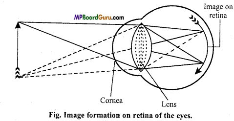
(b) Mechanism of vision: For seeing clear distant object it is essential that image must be focussed on the retina so that photoreceptor rods and cones are stimulated. Rays of light from the object fall on the biconvex lens after passing through the conjunctiva, cornea and pupil. The size of the pupil regulates the amount of light entering the eyeball. Thus, an inverted image of the object is formed on the retina which is conveyed to the brain by the optic nerve. The image is then reinverted by the brain.
The eyes have got the property to form image of both far and near objects on retina by changing the convexity of the lens. This is called power of accommodation.
(c) Mechanism of hearing: The sound waves first strike the pinna which keeps on moving to direct the sound waves into tympanic cavity. These waves then strike the tympanic membrane which starts vibrating due to which the ear ossicles are vibrated.
Thus it is transmitted the membrane over the fenestra ovalis into scala vestibule of the cochlea. In this way, the perilymph filled in it also gets vibrated due to which Reissner’s and basilar membranes start vibrating. By the vibration of these membranes, the endolymph of scala media also vibrates as well as the tectorial membrane.
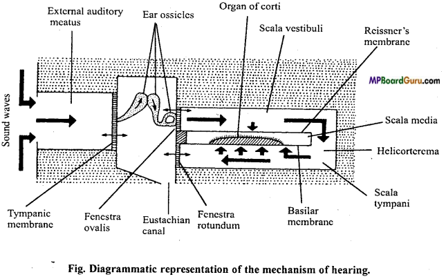
Because of the vibration of endolymph of scala media, the sensory hairs of the receptor cells of the organ of Corti are stretched and bent due to which the receptor cells gets stimulated and send the impulses to the brain through cochlear nerve. In the manner the sound is heard.
![]()
Question 7.
(a) Decribe role of Na+ in production of action potential.
(b) Describe role of Ca++ in discharge of neurotransmitter at svnapse.
(c) Explain mechanism of production of impulse on the retina by light.
(d) Explain mechanism of production of nerve impulse by sound in the internal ear.
Answer:
(a)
Nerve impulse: The sum total of the physical and chemical reactions which take place in the propagation of the wave of physiological activity along the nerve fibre is called as nerve impulse.
Propagation of Nerve impulse :
1. Depolarization: When a stimulus of any kind electrical, mechanical or chemical acts on the nerve fibre momentarily, a local increase occurs in the permeability of membrane by opening sodium gate at the site of stimulus. This suddenly brings sodium ions influx into the cell, carrying enough positive charges to the inside to cause complete disappearance of the normal resting potential and usually enough charges to develop a positive state inside the fibre. This positive state of the fibre is called reverse potential. It results in the depolarization of the membrane.
2. Action potential: The changed electrical potential of the plasma membrane of neuron is called action potential. The initial change produces an ionic imbalance across the membrane on either side of the points of stimulus producing local electrical current. These areas of negative depolarization in turn initiate changes in the membrane adjacent to them. A wave of electric charges or depolarization along the length of the nerve fibre or axon is called nerve impulse.
3. Repolarization: With the increase of positive charge inside further entry of Na+ is prevented and the permeability of membrane decreases. Na+ ions are pushed out which transfer large number of positive charge outside, once again creating negativity inside the membrane which restores the original resting potential. This is known as repolarization. The repolarization starts exactly on the same spot where depolarization had started and then continues to advance ahead. The entire process of repolarization requires some time during which the nerve cannot be stimulated again. It is known as refractory period.
Theories of nerve impulse conduction or Transmission :
1. Saltatory conduction or through nerve fibre : Myelin sheath acts as insulator in the medullated nerve fibres. It is almost impermeable to the ions. The membrane at the nodes of Ranvier is 500 times more permeable. The nerve impulse in these fibres passes from node to node along the entire length of nerve fibre rather than continuously along the entire fibre as occurs in unmyelinated fibre.
This process is called saltatory conduction. It is of dual value. First by causing the depolarization process to jump along nodes of Ranvier on the axis of the nerve fibre increases greatly the speed of impulse travel and secondly saltatory conduction conserves energy for the axon because only the nodes depolarize.
The velocity of conduction in nerve fibre varies from 5 metres per second in a small unmyelinated fibres to as high as 130 metres per second in large myelinated fibres.
Transmission of impulse across synapse (synaptic transmission): The area where the dendrite of one neuron is in close proximity with the axon of another is known as the synapse. The space between the axon and dendrite is called the synaptic cleft.
When the impulse reaches the tip of axon it stimulates the synaptic vehicles which move towards the synaptic cleft and discharge the neurotransmitter or neurohormones.
They then return the knob and become recharged. This chemical neurotransmitter substance has been identified as acetylcholine. Once released the neurotransmitter diffuses across the synaptic cleft and becomes attached to chemo- receptors present on the dendrites of the next neuron.
This attachment triggers the depolarization of the membrane and initiates action potential which travels across the next neuron. Thus, the actual transmission of the impulse across the synapse is by means of specific chemicals. Just after the transmission of impulse through the synapse, acetylcholine is hydrolyzed into acetic-acid and choline by the enzyme acetylcholinesterase present in the post synaptic membrane. The membrane again becomes polarized, so another impulse must come down the axon and release more acetylcholine for the transmission of another impulse.
(b)
(a)
Nerve impulse: The sum total of the physical and chemical reactions which take place in the propagation of the wave of physiological activity along the nerve fibre is called as nerve impulse.
Propagation of Nerve impulse :
1. Depolarization: When a stimulus of any kind electrical, mechanical or chemical acts on the nerve fibre momentarily, a local increase occurs in the permeability of membrane by opening sodium gate at the site of stimulus. This suddenly brings sodium ions influx into the cell, carrying enough positive charges to the inside to cause complete disappearance of the normal resting potential and usually enough charges to develop a positive state inside the fibre. This positive state of the fibre is called reverse potential. It results in the depolarization of the membrane.
2. Action potential: The changed electrical potential of the plasma membrane of neuron is called action potential. The initial change produces an ionic imbalance across the membrane on either side of the points of stimulus producing local electrical current. These areas of negative depolarization in turn initiate changes in the membrane adjacent to them. A wave of electric charges or depolarization along the length of the nerve fibre or axon is called nerve impulse.
3. Repolarization: With the increase of positive charge inside further entry of Na+ is prevented and the permeability of membrane decreases. Na+ ions are pushed out which transfer large number of positive charge outside, once again creating negativity inside the membrane which restores the original resting potential. This is known as repolarization. The repolarization starts exactly on the same spot where depolarization had started and then continues to advance ahead. The entire process of repolarization requires some time during which the nerve cannot be stimulated again. It is known as refractory period.
Theories of nerve impulse conduction or Transmission :
1. Saltatory conduction or through nerve fibre : Myelin sheath acts as insulator in the medullated nerve fibres. It is almost impermeable to the ions. The membrane at the nodes of Ranvier is 500 times more permeable. The nerve impulse in these fibres passes from node to node along the entire length of nerve fibre rather than continuously along the entire fibre as occurs in unmyelinated fibre.
This process is called saltatory conduction. It is of dual value. First by causing the depolarization process to jump along nodes of Ranvier on the axis of the nerve fibre increases greatly the speed of impulse travel and secondly saltatory conduction conserves energy for the axon because only the nodes depolarize.
The velocity of conduction in nerve fibre varies from 5 metres per second in a small unmyelinated fibres to as high as 130 metres per second in large myelinated fibres.
Transmission of impulse across synapse (synaptic transmission): The area where the dendrite of one neuron is in close proximity with the axon of another is known as the synapse. The space between the axon and dendrite is called the synaptic cleft.
When the impulse reaches the tip of axon it stimulates the synaptic vehicles which move towards the synaptic cleft and discharge the neurotransmitter or neurohormones.
They then return the knob and become recharged. This chemical neurotransmitter substance has been identified as acetylcholine. Once released the neurotransmitter diffuses across the synaptic cleft and becomes attached to chemo- receptors present on the dendrites of the next neuron.
This attachment triggers the depolarization of the membrane and initiates action potential which travels across the next neuron. Thus, the actual transmission of the impulse across the synapse is by means of specific chemicals. Just after the transmission of impulse through the synapse, acetylcholine is hydrolyzed into acetic-acid and choline by the enzyme acetylcholinesterase present in the post synaptic membrane. The membrane again becomes polarized, so another impulse must come down the axon and release more acetylcholine for the transmission of another impulse.
(c)
(a) Mechanism of synaptic conduction:
Nerve impulse: The sum total of the physical and chemical reactions which take place in the propagation of the wave of physiological activity along the nerve fibre is called as nerve impulse.
Propagation of Nerve impulse :
1. Depolarization: When a stimulus of any kind electrical, mechanical or chemical acts on the nerve fibre momentarily, a local increase occurs in the permeability of membrane by opening sodium gate at the site of stimulus. This suddenly brings sodium ions influx into the cell, carrying enough positive charges to the inside to cause complete disappearance of the normal resting potential and usually enough charges to develop a positive state inside the fibre. This positive state of the fibre is called reverse potential. It results in the depolarization of the membrane.
2. Action potential: The changed electrical potential of the plasma membrane of neuron is called action potential. The initial change produces an ionic imbalance across the membrane on either side of the points of stimulus producing local electrical current. These areas of negative depolarization in turn initiate changes in the membrane adjacent to them. A wave of electric charges or depolarization along the length of the nerve fibre or axon is called nerve impulse.
3. Repolarization: With the increase of positive charge inside further entry of Na+ is prevented and the permeability of membrane decreases. Na+ ions are pushed out which transfer large number of positive charge outside, once again creating negativity inside the membrane which restores the original resting potential. This is known as repolarization. The repolarization starts exactly on the same spot where depolarization had started and then continues to advance ahead. The entire process of repolarization requires some time during which the nerve cannot be stimulated again. It is known as refractory period.
Theories of nerve impulse conduction or Transmission :
1. Saltatory conduction or through nerve fibre : Myelin sheath acts as insulator in the medullated nerve fibres. It is almost impermeable to the ions. The membrane at the nodes of Ranvier is 500 times more permeable. The nerve impulse in these fibres passes from node to node along the entire length of nerve fibre rather than continuously along the entire fibre as occurs in unmyelinated fibre.
This process is called saltatory conduction. It is of dual value. First by causing the depolarization process to jump along nodes of Ranvier on the axis of the nerve fibre increases greatly the speed of impulse travel and secondly saltatory conduction conserves energy for the axon because only the nodes depolarize.
The velocity of conduction in nerve fibre varies from 5 metres per second in a small unmyelinated fibres to as high as 130 metres per second in large myelinated fibres.
Transmission of impulse across synapse (synaptic transmission): The area where the dendrite of one neuron is in close proximity with the axon of another is known as the synapse. The space between the axon and dendrite is called the synaptic cleft.
When the impulse reaches the tip of axon it stimulates the synaptic vehicles which move towards the synaptic cleft and discharge the neurotransmitter or neurohormones.
They then return the knob and become recharged. This chemical neurotransmitter substance has been identified as acetylcholine. Once released the neurotransmitter diffuses across the synaptic cleft and becomes attached to chemo- receptors present on the dendrites of the next neuron.
This attachment triggers the depolarization of the membrane and initiates action potential which travels across the next neuron. Thus, the actual transmission of the impulse across the synapse is by means of specific chemicals. Just after the transmission of impulse through the synapse, acetylcholine is hydrolyzed into acetic-acid and choline by the enzyme acetylcholinesterase present in the post synaptic membrane. The membrane again becomes polarized, so another impulse must come down the axon and release more acetylcholine for the transmission of another impulse.
(b) Mechanism of vision: For seeing clear distant object it is essential that image must be focussed on the retina so that photoreceptor rods and cones are stimulated. Rays of light from the object fall on the biconvex lens after passing through the conjunctiva, cornea and pupil. The size of the pupil regulates the amount of light entering the eyeball. Thus, an inverted image of the object is formed on the retina which is conveyed to the brain by the optic nerve. The image is then reinverted by the brain.
The eyes have got the property to form image of both far and near objects on retina by changing the convexity of the lens. This is called power of accommodation.
(c) Mechanism of hearing: The sound waves first strike the pinna which keeps on moving to direct the sound waves into tympanic cavity. These waves then strike the tympanic membrane which starts vibrating due to which the ear ossicles are vibrated.
Thus it is transmitted the membrane over the fenestra ovalis into scala vestibule of the cochlea. In this way, the perilymph filled in it also gets vibrated due to which Reissner’s and basilar membranes start vibrating. By the vibration of these membranes, the endolymph of scala media also vibrates as well as the tectorial membrane.
Because of the vibration of endolymph of scala media, the sensory hairs of the receptor cells of the organ of Corti are stretched and bent due to which the receptor cells gets stimulated and send the impulses to the brain through cochlear nerve. In the manner the sound is heard.
(d)
(a) Mechanism of synaptic conduction:
Nerve impulse: The sum total of the physical and chemical reactions which take place in the propagation of the wave of physiological activity along the nerve fibre is called as nerve impulse.
Propagation of Nerve impulse :
1. Depolarization: When a stimulus of any kind electrical, mechanical or chemical acts on the nerve fibre momentarily, a local increase occurs in the permeability of membrane by opening sodium gate at the site of stimulus. This suddenly brings sodium ions influx into the cell, carrying enough positive charges to the inside to cause complete disappearance of the normal resting potential and usually enough charges to develop a positive state inside the fibre. This positive state of the fibre is called reverse potential. It results in the depolarization of the membrane.
2. Action potential: The changed electrical potential of the plasma membrane of neuron is called action potential. The initial change produces an ionic imbalance across the membrane on either side of the points of stimulus producing local electrical current. These areas of negative depolarization in turn initiate changes in the membrane adjacent to them. A wave of electric charges or depolarization along the length of the nerve fibre or axon is called nerve impulse.
3. Repolarization: With the increase of positive charge inside further entry of Na+ is prevented and the permeability of membrane decreases. Na+ ions are pushed out which transfer large number of positive charge outside, once again creating negativity inside the membrane which restores the original resting potential. This is known as repolarization. The repolarization starts exactly on the same spot where depolarization had started and then continues to advance ahead. The entire process of repolarization requires some time during which the nerve cannot be stimulated again. It is known as refractory period.
Theories of nerve impulse conduction or Transmission :
1. Saltatory conduction or through nerve fibre : Myelin sheath acts as insulator in the medullated nerve fibres. It is almost impermeable to the ions. The membrane at the nodes of Ranvier is 500 times more permeable. The nerve impulse in these fibres passes from node to node along the entire length of nerve fibre rather than continuously along the entire fibre as occurs in unmyelinated fibre.
This process is called saltatory conduction. It is of dual value. First by causing the depolarization process to jump along nodes of Ranvier on the axis of the nerve fibre increases greatly the speed of impulse travel and secondly saltatory conduction conserves energy for the axon because only the nodes depolarize.
The velocity of conduction in nerve fibre varies from 5 metres per second in a small unmyelinated fibres to as high as 130 metres per second in large myelinated fibres.
Transmission of impulse across synapse (synaptic transmission): The area where the dendrite of one neuron is in close proximity with the axon of another is known as the synapse. The space between the axon and dendrite is called the synaptic cleft.
When the impulse reaches the tip of axon it stimulates the synaptic vehicles which move towards the synaptic cleft and discharge the neurotransmitter or neurohormones.
They then return the knob and become recharged. This chemical neurotransmitter substance has been identified as acetylcholine. Once released the neurotransmitter diffuses across the synaptic cleft and becomes attached to chemo- receptors present on the dendrites of the next neuron.
This attachment triggers the depolarization of the membrane and initiates action potential which travels across the next neuron. Thus, the actual transmission of the impulse across the synapse is by means of specific chemicals. Just after the transmission of impulse through the synapse, acetylcholine is hydrolyzed into acetic-acid and choline by the enzyme acetylcholinesterase present in the post synaptic membrane. The membrane again becomes polarized, so another impulse must come down the axon and release more acetylcholine for the transmission of another impulse.
(b) Mechanism of vision: For seeing clear distant object it is essential that image must be focussed on the retina so that photoreceptor rods and cones are stimulated. Rays of light from the object fall on the biconvex lens after passing through the conjunctiva, cornea and pupil. The size of the pupil regulates the amount of light entering the eyeball. Thus, an inverted image of the object is formed on the retina which is conveyed to the brain by the optic nerve. The image is then reinverted by the brain.
The eyes have got the property to form image of both far and near objects on retina by changing the convexity of the lens. This is called power of accommodation.
(c) Mechanism of hearing: The sound waves first strike the pinna which keeps on moving to direct the sound waves into tympanic cavity. These waves then strike the tympanic membrane which starts vibrating due to which the ear ossicles are vibrated.
Thus it is transmitted the membrane over the fenestra ovalis into scala vestibule of the cochlea. In this way, the perilymph filled in it also gets vibrated due to which Reissner’s and basilar membranes start vibrating. By the vibration of these membranes, the endolymph of scala media also vibrates as well as the tectorial membrane.
Because of the vibration of endolymph of scala media, the sensory hairs of the receptor cells of the organ of Corti are stretched and bent due to which the receptor cells gets stimulated and send the impulses to the brain through cochlear nerve. In the manner the sound is heard.
Question 8.
(a) How do you see colour of any object?
(b) Which part of our body helps to maintain equilibrium (balance) of the body?
(c) How does eyes regulate light which falls into the retina.
Answer:
(a) Retina contain two types of light receptor cells: Rods and cones. In the presence of light rods perceive light, whereas the cones are colour receptors, containing adoption or visual violet. They function in strong light and are meant for scotopic vision (colour vision), that is why we can not see colour in dim light or dark.
On the basis of pigments there are three types of cones :
- Cones respond to red light due to presence of erythroblast pigment.
- Cones respond to green light due to presence of chlorolabe pigment.
- Cones respond to blue light due to presence of cyanolabe pigment.
The colour that we see results from a mixture of impulses of the above types of cone
types.
(b) Ears play an important role in hearing and the maintenance of equilibrium and orientation of body, hence are called as balancing organ. Sensory cells are present in the ampullae of the semilunar canals and utriculus. Several particles of calcium carbonate or otoliths are scattered around the cilia of these cells. When the equilibrium of the body disturbed, the otoliths move and press upon the cilia or sensory cells which get stimulated and send the impulse to the brain. Thus brain receives the information and sends instructions to different organs to set right the equilibrium of body.
(c)
(a) Mechanism of synaptic conduction:
Nerve impulse: The sum total of the physical and chemical reactions which take place in the propagation of the wave of physiological activity along the nerve fibre is called as nerve impulse.
Propagation of Nerve impulse :
1. Depolarization: When a stimulus of any kind electrical, mechanical or chemical acts on the nerve fibre momentarily, a local increase occurs in the permeability of membrane by opening sodium gate at the site of stimulus. This suddenly brings sodium ions influx into the cell, carrying enough positive charges to the inside to cause complete disappearance of the normal resting potential and usually enough charges to develop a positive state inside the fibre. This positive state of the fibre is called reverse potential. It results in the depolarization of the membrane.
2. Action potential: The changed electrical potential of the plasma membrane of neuron is called action potential. The initial change produces an ionic imbalance across the membrane on either side of the points of stimulus producing local electrical current. These areas of negative depolarization in turn initiate changes in the membrane adjacent to them. A wave of electric charges or depolarization along the length of the nerve fibre or axon is called nerve impulse.
3. Repolarization: With the increase of positive charge inside further entry of Na+ is prevented and the permeability of membrane decreases. Na+ ions are pushed out which transfer large number of positive charge outside, once again creating negativity inside the membrane which restores the original resting potential. This is known as repolarization. The repolarization starts exactly on the same spot where depolarization had started and then continues to advance ahead. The entire process of repolarization requires some time during which the nerve cannot be stimulated again. It is known as refractory period.
Theories of nerve impulse conduction or Transmission :
1. Saltatory conduction or through nerve fibre : Myelin sheath acts as insulator in the medullated nerve fibres. It is almost impermeable to the ions. The membrane at the nodes of Ranvier is 500 times more permeable. The nerve impulse in these fibres passes from node to node along the entire length of nerve fibre rather than continuously along the entire fibre as occurs in unmyelinated fibre.
This process is called saltatory conduction. It is of dual value. First by causing the depolarization process to jump along nodes of Ranvier on the axis of the nerve fibre increases greatly the speed of impulse travel and secondly saltatory conduction conserves energy for the axon because only the nodes depolarize.
The velocity of conduction in nerve fibre varies from 5 metres per second in a small unmyelinated fibres to as high as 130 metres per second in large myelinated fibres.
Transmission of impulse across synapse (synaptic transmission): The area where the dendrite of one neuron is in close proximity with the axon of another is known as the synapse. The space between the axon and dendrite is called the synaptic cleft.
When the impulse reaches the tip of axon it stimulates the synaptic vehicles which move towards the synaptic cleft and discharge the neurotransmitter or neurohormones.
They then return the knob and become recharged. This chemical neurotransmitter substance has been identified as acetylcholine. Once released the neurotransmitter diffuses across the synaptic cleft and becomes attached to chemo- receptors present on the dendrites of the next neuron.
This attachment triggers the depolarization of the membrane and initiates action potential which travels across the next neuron. Thus, the actual transmission of the impulse across the synapse is by means of specific chemicals. Just after the transmission of impulse through the synapse, acetylcholine is hydrolyzed into acetic-acid and choline by the enzyme acetylcholinesterase present in the post synaptic membrane. The membrane again becomes polarized, so another impulse must come down the axon and release more acetylcholine for the transmission of another impulse.
(b) Mechanism of vision: For seeing clear distant object it is essential that image must be focussed on the retina so that photoreceptor rods and cones are stimulated. Rays of light from the object fall on the biconvex lens after passing through the conjunctiva, cornea and pupil. The size of the pupil regulates the amount of light entering the eyeball. Thus, an inverted image of the object is formed on the retina which is conveyed to the brain by the optic nerve. The image is then reinverted by the brain.
The eyes have got the property to form image of both far and near objects on retina by changing the convexity of the lens. This is called power of accommodation.
(c) Mechanism of hearing: The sound waves first strike the pinna which keeps on moving to direct the sound waves into tympanic cavity. These waves then strike the tympanic membrane which starts vibrating due to which the ear ossicles are vibrated.
Thus it is transmitted the membrane over the fenestra ovalis into scala vestibule of the cochlea. In this way, the perilymph filled in it also gets vibrated due to which Reissner’s and basilar membranes start vibrating. By the vibration of these membranes, the endolymph of scala media also vibrates as well as the tectorial membrane.
Because of the vibration of endolymph of scala media, the sensory hairs of the receptor cells of the organ of Corti are stretched and bent due to which the receptor cells gets stimulated and send the impulses to the brain through cochlear nerve. In the manner the sound is heard.
![]()
Question 9.
Describe functions of various parts of brain.
Answer:
Functions of different parts of brain :
Human brain consists of three parts :
- Fore-brain,
- Mid-brain,
- Hind-brain.
Their functions are as follows :
1. Fore-brain:
(A) Cerebrum :
- It is the center of intelligence and reasoning.
- It is the centre of memory.
- It regulates feeling.
- Different area of this part is responsible for specific function.
(B) Olfactory lobe: It is the centre of reflex action.
(C) Diencephalon :
- It recognizes the sensation such as heat, cold and pain.
- It regulates metabolism of carbohydrates and fats.
- It regulates blood pressure.
- It also regulates hunger, thirst, sleep, temperature, tiredness, hate and satisfaction feeling by autonomic nervous system present in it.
2. Mid-brain : It helps to receive stimuli of vision and hearing. ‘
3. Hind-brain :
(A) Cerebellum :
- It regulates contraction and relaxation of muscles.
- Regulates balance of the body.
(B) Medulla oblongata :
- Regulates all involuntary functions such as heartbeat, rate of respiration and contraction of alimentary canal.
- It regulates swallowing, vomiting, sneezing etc.
Question 10.
Write differences between following :
(a) Myelinated and Non-myelinated nerve fibre.
(b) Dendrites and Axon.
(c) Rods and Cones.
(d) Thalamus and Hypothalamus.
(e) Cerebrum and Cerebellum.
Answer:
(a) Differences between Myelinated and Nonmyelinated nerve fibre
| Myelinated nerve fibre | Non-myelinated nerve fibre |
| 1. The nerve fibre in which myelin sheath is found called as myelinated nerve fibre. 2. This type of nerve fibres are found in the spinal and cranial nerves. | The nerve fibre in which myelin sheath is not found called as non-myelinated nerve fibre. This type of nerve fibres are found in the autonomous and somatic nerve fibres. |
(b) Differences between Dendrites and Axon
| Dendrites | Axon |
| 1. Smaller processes arise from the cyton of neurons are called as dendrons, whichare further divided into branches called as dendrites. 2. Dendrites are responsible for receiving impulse from the receptors and conduct it to the cyton. | It is the longest process of the neuron. It conducts impulses from the cyton to the nerve endings. |
(c) Differences between Rods and Cones
| Rods | Cones |
| 1. It consists of Rhodopsin pigment, which is formed by vitamin A. 2. It perceives light. 3. In less intensity of light white and dark brown colour can be identified. 4. Deficiency of rods or its pigments causes night blindness disease. | It consists of three different pigments. It differentiates colour. |
(d) Differences between Thalamus and Hypothalamus
| Thalamus | Hypothalamus |
| 1. It consists of two round masses of grey matter bulging in the third ventricle. 2. Thalamus serves as a relay centre for sensory and motor impulses from spinalcord and brain stem to various parts of the cerebrum. | It forms the floor of diocoel and consists of patches of grey matter of neurosensory cells in the white matter. Hypothalamus links nervous system with endocrine system. It regulate endocrine system by secretion of neurohormones. It regulates carbohydrates and fat metabolism, blood pressure, water balance, temperature, hunger, thirst and emotions. |
(e) Differences between Cerebrum and Cerebellum
| Cerebrum | Cerebellum |
| 1. It is part of fore brain. 2. It is divided into two lobes called as cerebral hemispheres by a fissure. 3. It is the centre of intelligence, memory and reasoning. | It is part of hind brain. It is narrow towards the back side and connected with medulla oblongata. It is related to involuntary functions and body equilibrium. |
Question 11.
Write differences between following. (NCERT)
(a) Afferent neuron and Efferent neuron.
(b) Impulse conduction in Myelinated and Non-myelinated nerve fibres.
(c) Yellow spot and Blind spot.
(d) Aqueous humour and Vitreous humour.
(e) Cranial nerves and Spinal nerves.
Answer:
(a) Differences between Afferent neurons and Efferent neurons
| Afferent Neuron | Efferent Neurons |
| 1. They are found in dorsal root of spinal nerve. 2. They conduct the sensory impulse from receptor organ to central nervous system. 3. It is called as sensory nerve. | They are found in ventral root of spinal nerve. It passes the impulse from central nervous system to the effector organ. It is called as motor nerve. |
(b) Differences between Impulse conduction in Myelinated and Nonmyelinated nerve fibres
| Impulse conduction through Myelinated nerve fibres | Impulse conduction through Non-myelinated nerve fibres |
| 1. Myelinated nerve fibre is impermeable to ions. 2. The velocity of conduction in nerve fibre is 130meters per second 3. The nerve impulse in these fibres 4. Depolarisation occurs at the node. | Non-myelinated nerve fibre is permeable to ions. The velocity of conduction in nerve fibre is 5 meters per second. Conduction of impulse occurs along the entire fibre. Depolarisation occurs along the entire length of the member of the nerve fibre. |
(c) Differences between Yellow spot and Blind spot
| Yellow spot | Blind spot |
| 1. Posterior part of retina has yellow pigment. It is called yellow spot. 2. In yellow spot, rod and cones are present. | There is a point in retina where the optic nerve fibre leave the retina. This point is called blind spot In blind spot, rod and cones are absent. |
(d) Differences between Aqueous humour and Vitreous humour
| Aqueous humour | Vitreous humour |
| 1. Space between cornea and lens of the eye is called as Aqueous Chamber. 2. The aqueous chamber is filled with watery aqueous humour. | Space between lens and retina of the eye is called as vitreous chamber. The vitreous chamber is filled with gelatinous vitreous humour. |
(e) Differences between Cranial nerves and Spinal nerves
| Cranial nerves | Spinal nerves |
| 1. The nerves originating from brain are called as cranial nerves. 2. There are 12 pairs of cranial nerves. 3. Cranial nerves may be sensory, motor or mixed type in nature. | The nerves originate from spinalcord are called as spinal nerves. There are 31 pairs of spinal nerves. All spinal nerves are mixed nerves. |
Question 12.
How many cranial nerves are found in human? Give their name, nature, distribution and functions.
Answer:
Cranial nerves: In human 12 pairs of cranial nerves are found. Their name, nature, distribution and functions are as follows:
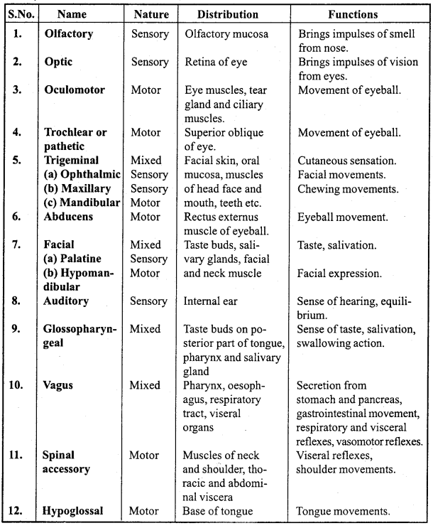
Question 13.
What is reflex action? Give its types. Describe mechanism of reflex action and give its importance.
Or,
What is reflex action? Draw well-labelled diagram of reflex action. Also explain the process of reflex action along with suitable example.
Answer:
Reflex action: The reflex actions are involuntary or unconscious response to a stimulus. They are brought about by reflex arc when a receptor is stimulated.
There are two types of reflexes :
1. Unconditional reflex: These are inborn reflexes and the nerve paths are the reflex arc fixed from the birth of the organism. They do not involve any thinking of brain, e.g. salivation of mouth, blinking of eye, knee jerk, etc. Complex in bom reflexes are called as instincts e.g. reproductive behaviour.
2. Conditional reflex: These are not inborn, but they can be acquired through practice or past experience.
Ventral fissure
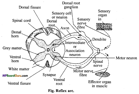
Mechanism of reflex action: A reflex arc comprises of some specific receptors, afferent neurons, connector or intermediate neurons, efferent neuron and effector organ. The impulses from the receptors pass on to the CNS through the afferent fibres whose axons terminate in the grey matter of either brain or spinal cord. In the grey matter these impulses are transferred to the motor neurons to carry out the specific action directly without any association neuron or they may be passed through one or many intermediate neurons to the effector organ. If a synapse is formed between the afferent and efferent fibres without a intermediate neuron it is known as mono synaptic reflex arc.
When there are two synapses, one between the afferent fibre and intermediate neuron and the other between the intermediate neuron and efferent fibre, it is known as bisynaptic reflex arc. The involvement of more than two intermediate neurons form a polysynaptic reflex arc. The involvement of more than two intermediate neurons form a polysynaptic reflex arc.
Some simple reflex actions:
1. Knee jerk: When patellar tendon is tapped, a quick jerk of the leg occurs.
2. Coughing or sneezing all of a sudden.
3. Withdrawal of hand or leg when it is pricked with a needle.
4. Blinking of eyelids when some object approaches the eye.
5. The quick recovery of balance of body after a sleep.
Importance of reflex action:
1. It takes less time as the process is regulated by spinal cord thus protect the body from accident.
2. Body of organism is protected all of a sudden.
3. Brain get more time to control other functions as reflex action is regulated by spinal cord.
Neural Control and Coordination Class 11 Important Questions Objective Type
1. Choose the correct answers:
Question 1.
Which of the following is the immediate covering of a nerve fibre :
(a) Sarcoplasm
(b) Perineurium
(c) Epineurium
(d) Endoneurium.
Answer:
(d) Endoneurium.
Question 2.
In humans, one of the following cranial nerves is associated with the sense of body balance:
(a) VI
(b) VII
(c) VIII
(d) IX.
Answer:
(c) VIII
![]()
Question 3.
If a person has lost his memory in an accident, the following part of the brain have got injured :
(a) Diencephalon
(b) Medulla oblongata
(c) Cerebellum
(d) Cerebrum.
Answer:
(d) Cerebrum.
Question 4.
Parasympathetic nervous system :
(a) Increases heartbeat
(b) Decreases heartbeat
(c) Originates heartbeat
(d) Has no effect upon heartbeat.
Answer:
(b) Decreases heartbeat
Question 5.
Brain is:
(a) Ectodermal
(b) Mesodermal
(c) Endodermal
(d) Mesoendodermal.
Answer:
(a) Ectodermal
Question 6.
The nervous strip connecting both the cerebral hemispheres in the rabbit is:
(a) Corpus callosum
(b) Corpus Albicans
(c) Corpus striatum
(d) Corpus spongiosum.
Answer:
(a) Corpus callosum
Question 7.
The thermoregulatory centre is situated in :
(a) Spinal cord
(b) Pituitary body
(c) Cerebellum
(d) Hypothalamus.
Answer:
(d) Hypothalamus.
![]()
Question 8.
The neurotransmitter which communicates between two neurons or between a neuron and a muscle is :
(a) Acetylcholine
(b) Globulin
(c) Rennin
(d) ATP.
Answer:
(a) Acetylcholine
Question 9.
What is found in the periphery of spinal cord :
(a) Grey matter
(b) Myelinated nerve
(c) White matter
(d) Notochord.
Answer:
(c) White matter
Question 10.
Which is correct about human brain :
(a) It is covered by two membranes
(b) There is no blood-brain barrier
(c) Largest number of cranial nerves originate from cerebral hemisphere
(d) Cerebral cortex is highly developed.
Answer:
(d) Cerebral cortex is highly developed.
Question 11.
Heart is innervated by :
(a) Trigeminal
(b) Vagus
(c) Glossopharyngeal
(d) Facial.
Answer:
(b) Vagus
Question 12.
Parotid gland located at the base of ear secretes :
(a) Wax
(b) Saliva
(c) Hormone
(d) Enzyme.
Answer:
(a) Wax
![]()
Question 13.
Tongue is controlled by :
(a) Trigeminal nerve
(b) Facial nerve
(c) Glossopharyngeal nerve
(d) None of the above.
Answer:
(b) Facial nerve
(c) Glossopharyngeal nerve
Question 14.
The co-ordination system of the body is :
(a) Blood vascular system
(b) Nervous system
(c) Endocrine system
(d) Nervous and Endocrine system.
Answer:
(d) Nervous and Endocrine system.
Question 15.
Synapse occurs in between :
(a) Nerve and muscles
(b) Two nerves
(c) Two nerve fibres
(d) One nerve fibre and neuron.
Answer:
(b) Two nerves
![]()
Question 16.
Which part out of the following is found covered with fatty covering :
(a) Axon
(b) Cyton
(c) Dendrite
(d) Node of Ranvier.
Answer:
(a) Axon
Question 17.
Sequence of ear ossicles is :
(a) Incus Malleus stapes
(b) Stapes incus malleus
(c) Malleus incus stapes
(d) None of the above.
Answer:
(c) Malleus incus stapes
Question 18.
Part of the eye of vertebrate from where optic nerve comes out: (NCERT)
(a) Phobia
(b) Iris
(c) Blind spot
(d) Optic chiasma.
Answer:
(c) Blind spot
2. Fill in the blanks:
1. The junction present between two neurons is called ……………… .
Answer:
Synapsis,
2. Acetyl, choline is a ……………… .
Answer:
Neurotransmitter,
3. In human being ……………… pairs of spinal nerves and ……………… pairs of cranial nerves are found.
Answer:
31, 12,
4 ……………… is originated from optic nerve.
Answer:
Blindspot,
5. The cones of light are sensitive to only ……………… light.
Answer:
Bright,
6. All the nerves coming out of the spinal cord are ……………… nerves.
Answer:
Mixed,
7. Internal ear is made up of ……………… labirynth and ……………… labirynth.
Answer:
Bony, Membranous,
8. The eyes of nocturnal animals contain only ……………… .
Answer:
Rods,
9. Eustachian tube is found in the ……………… .
Answer:
Middle ear,
10. Corpus callosum is found in the ……………… .
Answer:
Human brain
![]()
3. Match the following:
(A)
| Column ‘A’ | Column ‘B’ |
| 1. Meninges | (a) Respiratory centre |
| 2. Organs of corti | (b) Reflex action |
| 3. Cerebralhemisphere | (c) Brain |
| 4. Spinal cord | (d) Choclea |
| 5. Medulla oblongata | (e) Thinking. |
Answer:
1. (c) Brain,
2. (d) Choclea
3. (e) Thinking.
4. (b) Reflex action
5. (a) Respiratory centre
(B)
| Column ‘A’ | Column ‘B’ |
| 1. Statolith | (a) Colour |
| 2. Cone | (b) Internal ear |
| 3. Jacobson organ | (c) Balance |
| 4. Endolymph | (d) Duramater |
| 5. Brain | (e) Smell. |
Answer:
1. (c) Balance
2. (a) Colour
3. (e) Smell.
4. (b) Internal ear
5. (d) Duramater.
4. Write true or false :
1. Name the part of retina which contains only cones.
Answer:
Yellow spot,
2. Give an example of general reflex action.
Answer:
Blinking of eyes,
3. Write the name of fluid present in eye.
Answer:
Aqueous and vitreous humous,
4. Write the name of nerve responsible for the movement of tongue.
Answer:
Hypoglacel nerve,
5. Write the name of smallest bone of human ear.
Answer:
Stapes,
6. Write the number of cranial and spinal nerves found in a human.
Answer:
12 pairs, 31 pairs,
7. Name the pigment which is helpful in vision during night.
Answer:
Rhodopsin, [photopic vision (black and white)]
8. Name the pigment present in the skin.
Answer:
Melanin,
9. The black colour of elephant indicates which place it lives.
Answer:
9. Hot places.