Students get through the MP Board Class 11th Biology Important Questions Chapter 18 Body Fluids and Circulation which are most likely to be asked in the exam.
MP Board Class 11th Biology Important Questions Chapter 18 Body Fluids and Circulation
Body Fluids and Circulation Class 11 Important Questions Very Short Answer Type
Question 1.
Which type of circulation is found in unicellular organisms?
Answer:
Unicellular organisms shows intracellular circulation. Cytoplasm of cell of the unicellular organism shows streaming movement called cyclosis. It helps in the circulation and distribution of materials in the cells e.g. Amoeba, Paramoecium etc.
Question 2.
What is known as heart block?
Answer:
Heart block : It is a condition in which bundle of His’ does not function properly then heartbeating impulse of atrial node does not reach ventricles, thus ventricle cannot exhibit their movement and thus circulation of blood is inhibited. This condition is called as heart block.
![]()
Question 3.
Where does the ‘Lubb’ and ‘Dup’ sounds are produced in Cardiac cycle?
Answer:
Lubb: ‘Lubb’ sound is produced during closure of atrioventricular valve (Bicuspid and Tricuspid valves). It is the first heart sound, which is low pitched but are of longer duration.
Dup : It is produced during closure of semilunar valves present at the base of arterial trunks. It is the second heart sound which is high pitched but of shorter duration.
Question 4.
Give reason that arteries are thick walled than that of veins.
Answer:
Arteries are thick walled than that of veins: When heart contracts blood enters into the arteries with great pressure as heart is responsible for supply of blood to each part of the body. To exert this pressure arteries are thick walled, whereas veins carry blood from different parts of the body to the heart without any pressure thus they are thin walled.
Question 5.
Write two functions of pericardial fluid.
Answer:
Two main functions of pericardial fluid are :
(i) It protects the heart from outer injuries,
(ii) It protects the heart from dryness.
Question 6.
Human heart is called myogenic. Why? (NCERT)
Answer:
Myogenic hearts are those where waves of contraction originate in special muscle fibres of heart.
Question 7.
What do you understand by double circulatory system? What is its significance? (NCERT)
Answer:
Double circulatory system: The circulatory system in which blood passes twice through heart for completing one cycle is called double circulatory system. In this type of circulatory system, oxygenated and deoxygenated blood flow remain separate. This type of circulation is found in all mammalian animals. In this system, first the deoxygenated blood is collected by veins into the heart and then it goes to the lungs for oxygenation. It returns to the left part of the heart and then to the body organs through arteries. Thus, the blood has to travel twice in the heart before it circulates in the body.
Question 8.
What is carboxyhaemoglobin?
Answer:
P-wave It is complex formed by the combination of haemoglobin with the CO2. It provides blue colour to the blood in the vein.
Hb + CO2 → Hbc
![]()
Question 9.
What are thrombocytes ? Where do they occur?
Answer:
In human blood, some irregular shaped cells are present which help in clotting of blood are called thrombocytes. They are formed in bone marrow.
Question 10.
In blood vessels, the flowing blood do not clot, why?
Answer:
In blood vessels, the flowing blood do not clot because in blood plasma, antiprothromb in, heparin named carbohydrate is present which takes the active thrombin to passive prothrombin direction due to which vessels do not clot.
Question 11.
What is pulse? What is the pulse rate of human beings?
Answer:
Beating of the heart is also felt in the surface arteries as regular jerk called as pulse. Each ventricular systole starts a new pulse, which proceeds as a wave of expansion through arteries, which is repeated after every 08 seconds.
It can be felt at the radial artery of wrist, temporal artery in front of the ear and carotid artery of the neck etc. Pulse rate of normal adult is 72 times per minute. In children, it is 120 times per minute and in old persons it is 60 times per minute.
Question 12.
How does partial pressure of respiratory gases control diffusion of oxygen from blood capillaries to the tissue?
Answer:
In the body tissue, P02 is very low that is about 40mm Hg, hence oxygen from arterial blood where P02 is about 95mm Hg. Thus oxygen separates from haemoglobin and diffuses into the tissues until the P02 goes down to 40mm Hg.
Question 13.
How does regulation of heartbeat occurs?
Answer:
The normal rate of heart beat is regulated by the following three ways :
- Nervous control: Cardiac centre lies in the medulla of brain regulate heartbeat.
- Hormonal control: Adrenalin and Noradrenalin hormone increases and decreases heart activity respectively.
- Chemical control: Vagus nerve on stimulation secretes acetyl-choline which slows down the heartbeat while tip end of sympathetic nerve fibre secretes adrenalin, which accelerates heartbeat.
Question 14.
What is Heart sound?
Answer:
Various valves present at the inlet and outlet of ventricles regulate the flow of blood in the heart. Action of these valves, while closing the apertures, create the heart sound. Each heartbeat is accompanied by two heart sound ‘Lubb’ and ‘Dup’. It can be heard using a device called as stethoscope.
Question 15.
What is haemocoel?
Answer:
In higher invertebrates like arthropods and molluscs, open circulatory system is found. Blood vessels of them*are not divided into capillaries and open into write spaces called as sinuses in the body cavity, the blood fills in the body cavity and baths the organs. The body cavity filled with blood is called as haemocoel and the blood which is colourless called as haemolymph.
Body Fluids and Circulation Class 11 Important Questions Short Answer Type
Question 1.
Explain heart sound. (NCERT)
Answer:
Refer Q. No. 3 and 14 of Very Short Answer Type Questions.
Question 2.
Define Cardiac cycle and Cardiac output. (NCERT)
Answer:
Cardiac cycle: Contraction of the heart is called as Systole and relaxation of heart is called as Diastole. The sequence of one systole followed by one diastole is known as Cardiac cycle. It takes 0-8sec time to complete one cardiac cycle.
Cardiac output: At each heartbeat ventricle pump about 70ml blood. This volume is called as stroke volume. The heart beats 72 times per minute, which is known as heart rate. Quantity of blood pumped by the heart in one minute is called as cardiac output, i. e. 72 x 70ml = 5040ml blood per minute.
![]()
Question 3.
Where bicuspid, tricuspid and semilunar valves are present in human heart ? Write its functions.
Answer:
1. Bicuspid valve: The atrioventricular opening between left auricle and left ventricle is guarded by two flaps known as Bicuspid valve. It prevents backflow of blood.
2. Tricuspid valve: The right atrioventricular opening is guarded by three flaps known as tricuspid valve.
3. Semilunar valve: These are half-moon shaped flaps arising from the base of the pulmonary artery and aorta. A set of three semilunar valves guard the opening of right ventricle into pulmonary artery and another set is present where aorta arises from the left ventricle. All these valves present backflow of blood into ventricles.
Question 4.
Why blood is transported from auricles to ventricles during the relaxation of ventricles or ventricular diastole ?
Answer:
The volume of the ventricles is increased during the relaxation of ventricles and the inside pressure of it is low in comparison to the pressure of arteries. This will cause the closure of semilunar valves. Ventricles are remain in the position of relaxation even after closing of these semilunar valves.
Thus, lower pressure of their wall is maintained as it is. To balance this pressure aurio-ventricular valves are opened and blood flows from auricles to ventricles and fill up their cavity. After then ventricles contracts and thus the blood of its cavity is transported into arteries. Both of these processes (contraction and relaxation) occur alternatively.
![]()
Question 5.
What do you understand by blood pressure? Explain systolic and diastolic blood pressure.
Answer:
The pressure exerted on blood capillaries by pumping action of heart or by circulating blood of the body is called blood pressure.
Or
The force or pressure which the blood exerts on the walls of the artery in which it is contained is known as blood pressure.
Systolic pressure: Due to contraction of heart, blood is thrown out from the heart into artery which causes maximum blood pressure. This maximum pressure of blood in the blood capillary is called systolic blood pressure. The systolic pressure of a person is 120 mm Hg.
Diastolic pressure: The blood pressure of biood capillaries is minimum during the relaxation of ventricles. The minimum blood pressure of blood capillaries is called diastolic pressure. The diastolic blood pressure of an adult person is 80 mm Hg.
Question 6.
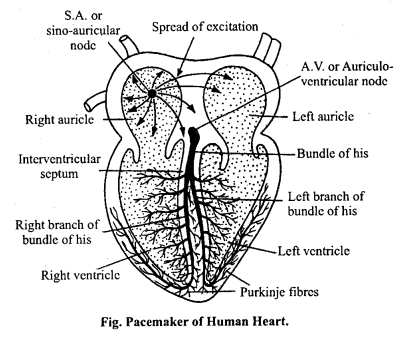
Where is sinoauricular node (SAN) situated? Why is SA node called pacemaker (speed motivator) of heart? (NCERT)
Answer:
Sinoauricular node (SA node) is situated in the upper part of right auricle of the heart. It is the site where heartbeat originates. It is also called as pacemaker or pacesetter. It is called pacemaker of the heart because cardiac rhythms commence here. When SA node does not function accurately the rate of heart beating will be irregular, slow or it stops. In this condition heart can not pump the blood according to requirement. In this condition artificial cardiac pacemaker is the substitute for a natural defective pacemaker which controls the beating of heart. Thus, SA node determines the rate of heartbeat and initiates it and so it is called as pacemaker of the heart.
Question 7.
Describe any four differences between sinoauricular and auriculo-ven- tricular node.
Answer:
Differences between S.A. Node and A.V. Node
| S. A. Node | A.V. Node |
| 1. It is located in the right auricles. | It is located in the grooves where the auricles and the ventricles meet together. |
| 2. It is the pacesetter of heart. | It is partially helping in the pace setting of heart. |
| 3. It is the area of origin of heart beat. | It is the second area of transmitting heartbeat. |
| 4. Accessory branches are not associated with S.A. node. | Two additional branches are associated with A.V. node. |
Question 8.
Write differences between the following : (NCERT)
(a) Closed and Open Circulatory system
(b) Mitral value and Semilunar value.
Answer:
(a) Differences between Closed and Open circulatory system
| Closed circulatory system | Open circulatory system |
| 1. In this type of circulatory system blood does not pass into open space but is always contained inside blood vessels e.g., Earthworm. | In this type of circulatory system blood passes into open space surrounding the living cells e.g., Cockroach. |
| 2. In this type of circulatory system, blood does not fill the body cavity. | In this type of circulatory system, the blood fills body cavity, called haemocoel. |
| 3. In this type of circulatory system, the blood flows with a pressure. e.g. It is found in vertebrates. | Blood does not flow with pressure. e.g. It is found in insects. |
(b) Differences between Mitral value and Semilunar value
| Mitral value | Semilunar value |
| 1. It is located at the left auriculoven tricular aperture. | It is located at the opening of ventricles into the aortas. |
| 2. It prevent backword flow of blood from left ventricle to left auricle. | It prevent backward flow of blood from aortas to ventricles. |
| 3. It closes the aperture by making a sound ‘Lubb’. | It closes the aperture by making a sound ‘Dup’. |
Question 9.
Write any four differences between an Artery and a Vein.
Answer:
Differences between an Artery and a Vein
| Artery | Vein |
| 1. Arteries transport blood from heart to the other organs of the body. | Veins collect the blood from various organs of the body into heart. |
| 2. Arteries contain oxidized blood. (except pulmonary artery) | Veins contain deoxidized blood. (except pulmonary vein). |
| 3. The wall of artery is thick and elastic. | The wall of vein is thin. |
| 4. Arteries are dark red in colour because they contain oxidized blood (pure blood). | Veins are blue in colour because its blood contains CO2 |
Question 10.
What are open and closed vascular system? Give two significance of closed vascular system.
Or,
What is closed vascular system?
Answer:
Open vascular system: The circulatory system in which blood flows indepen-dently in the coelomic cavity is called as open circulatory system.
Example: Cockroach.
Closed vascular system: The circulatory system in which blood flows through closed vessels.
Example: Earthworm, human.
Significance of closed vascular system :
- Blood contains haemoglobin which helps to carry oxygen during respiration.
- It helps in fast removal of excretory substance from the body.
- Valves present in its blood vessels, prevents backward flow of blood.
Question 11.
Write differences between Blood and Lymph.
Answer:
Differences between Blood and Lymph
| Blood | Lymph |
| 1. It is a red coloured fluid. | It is a colourless fluid. |
| 2. R.B.Cs. are found in more number. | R.B.Cs. are absent. |
| 3. W.B.Cs. are less in number. | W.B.Cs. are more in number. |
| 4. Fibrinogen is more in number. | Fibrinogen is less in number. |
| 5. Protein is found in more number. | Protein is found in less number. |
| 6. Excretory substances are found in less quantity. | Excretory substances are found in more quantity. |
| 7. Quantity of O2 is more. | Quantity of O2 is less. |
| 8. Food materials are found in more quantity. | Food materials are found in less quantity. |
Question 12.
Give importance of Plasma protein. (NCERT)
Answer:
There are three plasma proteins :
- Serum globulin,
- Serum albumin,
- Fibrinogen.
Functions or Importance of Plasma protein :
1. Immunity of the body: Globulin protein acts as antibody thus provide immunity to disease.
2. Prevent loss of blood by blood clotting: Fibrinogen and prothrombin are essential for clotting of blood.
3. Maintain fluidity of blood: Albumin and globulin possesses the ability to retain water in the blood plasma by their osmotic effects.
4. It helps for transport of protein and other substances.
5. It helps to maintain the pH of the blood.
6. It helps to maintain constant temperature of the body.
7. It helps to conduct heat.
![]()
Question 13.
Describe various components of blood and give one function of each component. (NCERT)
Answer:
Various component of blood and their functions :
Blood: Blood is a liquid connective tissue. Its matrix is liquid called as plasma. 90% of plasma is water in which various inorganic and organic substances are found in dissolved form. Three types of cells are found embedded in plasma called as corpuscles :
1. Erythrocytes or Red Blood Corpuscles (R.B.Cs.): These are biconvex, disc shaped, anucleated cells which contains a pigment haemoglobin. It helps to carry O2 and CO2 during respiration.
2. Leucocytes or White Blood Corpuscles (W.B.Cs.): These are nucleated, colourless, amoeboid cells. They are found in less number. They are of two types: It prevents the body from diseases by digesting and killing pathogenic germs.
3. Blood platelets: These are small, colourless, flat, granular, non-nucleated corpuscles, which helps for clotting of blood at injured part of the body.
Question 14.
Why blood is considered as connective tissue?
Answer:
Group of cells which are similar in their structure, function and origin are called as tissue.
Structurally blood is similar to connective tissue as it has a liquid matrix called as plasma in which various types of blood cells, RBCs, WBCs and Blood platelets are found in scattered form. It is also functionally similar to connective tissue as it connects all parts of the body by transporting materials. Thus blood is called as liquid connective tissue.
Body Fluids and Circulation Class 11 Important Questions Long Answer Type
Question 1.
Write differences : (NCERT)
(a) Blood and Lymph
(b) Open and Closed Circulatory System
(c) Systole and Diastole
(d) P-wave and T-wave.
Answer:
(a) Differences between Blood and Lymph
| Blood | Lymph |
| 1. It is a red coloured fluid. | It is a colourless fluid. |
| 2. R.B.Cs. are found in more number. | R.B.Cs. are absent. |
| 3. W.B.Cs. are less in number. | W.B.Cs. are more in number. |
| 4. Fibrinogen is more in number. | Fibrinogen is less in number. |
| 5. Protein is found in more number. | Protein is found in less number. |
| 6. Excretory substances are found in less quantity. | Excretory substances are found in more quantity. |
| 7. Quantity of O2 is more. | Quantity of O2 is less. |
| 8. Food materials are found in more quantity. | Food materials are found in less quantity. |
(b)
(a) Differences between Closed and Open circulatory system
| Closed circulatory system | Open circulatory system |
| 1. In this type of circulatory system blood does not pass into open space but is always contained inside blood vessels e.g., Earthworm. | In this type of circulatory system blood passes into open space surrounding the living cells e.g., Cockroach. |
| 2. In this type of circulatory system, blood does not fill the body cavity. | In this type of circulatory system, the blood fills body cavity, called haemocoel. |
| 3. In this type of circulatory system, the blood flows with a pressure. e.g. It is found in vertebrates. | Blood does not flow with pressure. e.g. It is found in insects. |
(b) Differences between Mitral value and Semilunar value
| Mitral value | Semilunar value |
| 1. It is located at the left auriculoven tricular aperture. | It is located at the opening of ventricles into the aortas. |
| 2. It prevent backword flow of blood from left ventricle to left auricle. | It prevent backward flow of blood from aortas to ventricles. |
| 3. It closes the aperture by making a sound ‘Lubb’. | It closes the aperture by making a sound ‘Dup’. |
(c) Differences between Systole and Diastole
| Systole | Diastole |
| 1. The contraction phase of the heart is called as Systole. | The relaxation phase of the heart is called as Diastole. |
| 2. Systole of heart pump out the blood from heart to different parts of the the body. | Diastole of heart decreases pressure in heart, thus blood from all parts of the body enters into the heart. |
(d) Differences between P-wave and W-wave
| P-wave | W-wave |
| 1. P-wave indicates the impulse of contraction gene-rated by sinu-auricular node of the right auricle | T-wave represents the relaxation of ventricles. |
Question 2.
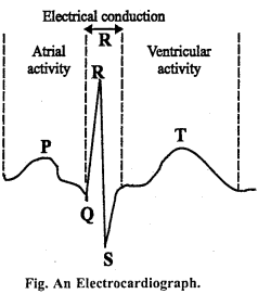
Draw a Unit of ECG and explain its various segments. (NCERT)
Answer:
Electrocardiogram (ECG): A graphic record of the electrical variations produced by the beating of the heart is called electrocardiograph. These variations are due to the development of electrical negativity of excited muscles as compared with unexcited tissues. An instrument used to observe the working of the heart is called electro-cardiogram. It was discovered by Einthoven (1906).
ECG is formed by a series of ridges and furrows. Normal pattern of ECG for a healthy person is given below.
In this, ‘P’ wave indicates the impulse of contraction generated by S.A. node. ‘QRS’ wave indicates the spread of impulse of contraction from A. V. node to the wall of ventricles, whereas the ‘T’ wave represents the relaxation of ventricles.
Any abnormality in the working of the heart changes the wave pattern of ECG and can be interpreted by a trained physician to diagnose a particular disorder. The technique to detect the abnormalities of the heart with the help of ECG is called electrocardiography.
Question 3.
Give reasons of following :
(a) Arteries are thick walled than that of veins.
(b) Doctors give injection in the veins.
(c) Veins are provided with valves but not arteries.
(d) Veins are blue coloured whereas arteries are red in colour.
Answer:
(a) Arteries are thick walled than that of veins: When heart contracts blood enters into the arteries with great pressure as heart is responsible for supply of blood to each part of the body. To exert this pressure arteries are thick walled, whereas veins carry blood from different parts of the body to the heart without any pressure thus they are thin walled.
(b) Doctors give injection in the veins: Doctors give injection in the veins because they are found in the surface of the body and carries blood to the heart, from where medicine can be distributed to all parts of the body through arteries.
But if doctors will give injection to the arteries then medicine will reach to particular part only.
(c) Veins are provided with valves but not arteries: Veins carries blood from dif-ferent part of the body to the heart without any pressure thus in order to prevent backward flow of blood and to direct the blood towards the heart, veins are provided with valves. Whereas arteries carry blood from heart to different parts of the body with pressure thus blood can easily reach to all parts of the body. Therefore arteries are not provided with valves.
(d) Veins are blue coloured whereas arteries are red in colour: Outer wall of the vein consists of tunica externa. Cells of this layer contain a pigment substance which reflect blue light thus veins appears blue whereas such pigments are not found in the wall of the arteries. Thus, arteries appears red coloured due to presence of blood in it, which reflect red light.
![]()
Question 4.
Give reasons of following statements :
(a) Wall of the left ventricle is more thick as compared to the wall of the right ventricle.
(b) Wall of the left ventricle is thick as compared to the auricles.
(c) Lymph contains less protein as compared to the plasma.
(d) Closed circulatory system is better as compared to the open circulatory system.
(e) During systole ventricles of the heart becomes a closed chamber.
Answer:
(a) Wall of the left ventricle is more thick as compared to the wall of the right ventricle : Right ventricles contains impure blood. To pass blood to the pulmonary artery less pressure is required by the right ventricle whereas to send blood to cortico sys-temic aorta more pressure is required by the left ventricle thus wall of the left ventricle is thicker than that of right ventricle.
(b) Wall of the left ventricle is thick as compared to the auricles : Left ventricle is responsible for the supply of blood to all parts of the body through cortico systemic aorta thus more pressure is required by it where as auricle receives blood without any pressure. Therefore left ventricle is thick walled as compared to the auricles.
(c) Lymph contains less protein as compared to the plasma : Capillaries found in the lymph are impermeable thus plasma protein quantity of lymph is less as compared to the blood.
(d) Closed circulatory system is better as compared to the open circulatory system:
In open circulatory system blood do not flow through closed vessels. Blood is found in the haemocoel cavity and is direct in contact of the organs. Blood pressure in this system is very less, e.g. Cockroach. In closed circulatory system blood is enclosed by heart, arteries and veins. Blood flows fast through these vessels and come back to the heart.
In this way exchange of substances between blood and tissue occurs fast. Whereas in open circulatory system this type of regulation is not found, thus closed circulatory system is considered as best as compared to the open circulatory system.
(e) During systole ventricles of the heart becomes a closed chamber : During ventricular systole pressure of the ventricle is more than that of auricles. Thus when blood reaches to the ventricle from auricles auriculoventricular valves closes the auriculo-ven- triculi aperture and prevents backward flow of blood. As ventricular pressure is less than the pressure of pulmonary artery thus semilunar valves remain closed, thus ventricle con-tracts as a closed chamber.
Question 5.
Who can donate blood to whom, also write the precautions of blood donation?
Answer:
Blood transfusion depends mainly on blood group. Therefore :
1. A blood group can be donated to person with blood group type A and AB.
2. Person with blood group B can donate blood to person with blood group type B or AB.
3. Person with AB group can donate blood only to person with AB blood group.
4. Blood group O is called universal donor but he can receive blood of only group O type.
Similarly the persons with blood group AB are called universal recipient as they can receive blood from persons with any blood group type.
Precautions of blood donation :
1. The first and the foremost precaution for donation of blood is testing the blood groups of both the donor and the recipient so that the problem of agglutination can be avoided.
2. Many fatal diseases get transmitted through blood. Thus, the donor’s blood should be tested for the presence of HIV and Hepatitis virus and getting negative result should be confirmed before transfusion.
3. Only upto 500 ml of blood should be donated by a man at one time.
4. Blood should not be donated more than twice in one year.
5. Donor should be completely healthy.
6. Equipments used in the process of transfusion should be thoroughly sterilized before use.
![]()
Question 6.
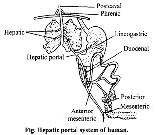
What is Hepatic portal system?
Answer:
Generally after collecting deoxygenated blood veins pour the blood into heart. But some veins pour blood into some other organs before reaching to heart for its purification. There veins are called as portal veins, and the system formed by these veins is called as portal system.
Hepatic portal system of man : When portal veins carry blood to the liver instead of heart and then through some other veins from liver to heart, it is called as Hepatic portal system.
Hepatic portal veins of human collect blood from different parts of the alimentary canal and carry it to liver. From liver through hepatic vein blood reaches to post caval.
Hepatic portal system consists of following veins :
- Lineogastric vein: It collects blood from all wall of the stomach and spleen.
- Duodenal veins: It collects blood from duodenum.
- Anterior mesenteric vein: It collects blood from small intestine, caecum and colon.
- Posterior mesenteric vein: It collects blood from rectum and anus.
Question 7.
Describe the structure of mammalian heart.
Or,
Describe structure and mechanism of working of mammalia heart.
Or,
Draw a well labelled diagram of L.S. of heart.
Answer:
Structure of Human Heart:
Position and external structure of human heart: The mammalian heart including man is a hollow, cone-shaped, muscular structure that lies in the thoracic cavity above the diaphragm and in between the two lungs. It is about the size of a fist measuring about 12 cm in length and 9 cm in breadth. It’s weight is about 300 grams. It is four-chambered organ, having two auricles and two ventricles.
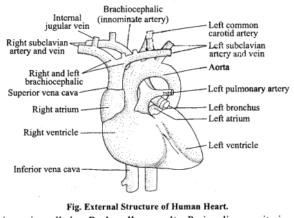
Heart lies in cavity called as Pericardium cavity. Pericardium cavity is surrounded by double membrane and is filled with pericardial fluid, which prevent the heart from external jerk, friction and keeps the heart moist.
Internal structure of human heart: The auricles are thin walled, sac like structures. The right and left auricles are separated from each other by a longitudinal muscular partition, called interauricular septum. The anterior and posterior vena cava open by separate apertures in the right auricles. The pulmonary veins open into left auricle by single common opening. The cavity of the ventricle is also divided into two by a thick and muscular interventricular septum. The wall of the left ventricle is much thicker than that of the right. The cavity of the left ventricle is circular while that of right is crescentic in outline in transverse sections. The right auricle opens into the right ventricle by a wide aperture called right auriculoventricular aperture. Similarly, the left auricle opens into the left ventricle by means of a large opening known as left auriculoventricular aperture.
The right auriculoventricular aperture is guarded by a valve which is formed of three flaps or cusps. This valve is called right auriculoventricular valve or tricuspid valve. The left auriculoventricular aperture is provided with a valve which has two cusps. It is known as left auriculoventricular valve or bicuspid valve or mitral valve. When the ventricles contract, both these valves close so that the blood is prevented from going back into the auricles from the ventricles. These valves are attached to columnae carnae or papillary muscles on the inner wall of the ventricles by means of thin threadlike structures called choradae tendinae.
The walls of the ventricles are much thicker than those of the auricles and are highly muscular. A special type of muscles are grouped together on the top of the interventricular septum to form a structure called Bundle of His or atrioventricular bundle. From the left anterior part of right ventricles arises the pulmonary arch which curves towards the left side of the heart passing below the carotid-systemic arch and goes to the lungs from the dorsal side of the heart.
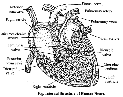
Three semilunar valves are present at the origin of the pulmonary arch. These valves do not allow the blood to go back into the right ventricles. The aorta or carotid-systemic arch arises from the right anterior part of the left ventricle. This arch passes over the pulmonary arch to come over the dorsal side of the heart where it forms the dorsal aorta below the vertebral column. These semilunar valves are also present at the origin of this arch to check the backflow of the blood into the left ventricle.
Mechanism of working of Human heart:
Mechanism of human heart: The rhythmic contraction and relaxation of the auricles and ventricles of heart in a specific sequence is called heart beat. The contraction phase is called as systole and the relaxation phase is called diastole.
The systole and the diastole together constitute a heart beat. The movement of the auricles and the ventricles are repeated in a cyclic manner during each heart beat. The amount of blood ejected by each ventricle per stroke at rest is about 70-90 ml. The volume of blood is termed as the stroke volume. This leaves about 50 ml of blood in each ventricle at the end of a systole and is known as end-systolic ventricular blood volume.
The human heart beats about 72 times a minute and this is termed as heart rate. The stroke volume when multiplied by the heart rate gives the volume of blood pumped out by each ventricle per minute. This volume is referred to as cardiac output.
Cardiac cycle: The atrial and ventricular systole and diastole which occur during each complete heart beat is called a cardiac cycle. This happens for a specific duration and the blood flows through the heart in a specific direction. The duration of each cardiac cycle at a heart rate of 75 per minute is 0-80 seconds in which the duration of systole is 0-27 seconds and that of diastole is 053 seconds.
Late diastole: In the late diastole the bicuspid and the tricuspid valves between the auricles and ventricles are opened and the semilunar valves at the entrance of aorta and pulmonary artery are closed. Blood flows into the heart throughout diastole, filling the auricles and ventricles. The rate of filling declines as the ventricles become distended and the cusps of the A.V. valves drift toward the closed position.
Atrial systole: Over two-thirds of the ventricular filling occurs passively during dias-tole. Contraction of the auricles propels some additional blood into the ventricles. The con-traction of the atria is called atrial systole. Contraction of the atrial muscles that surrounds the orifices of the vena cava and pulmonary veins blocks their opening and so the atrial blood cannot pass back from the atria into these vessels, but there is some regurgitation of blood into the veins during atrial systole.
Ventricular systole : The simultaneous contraction of both the ventricles is called ventricular systole. The initial portion of ventricular systole is called isometric or isovolumetric ventricular contraction which lasts until the aortic and pulmonary valves open. The A.V. valves close at the start of isometric contractions. Intraventricular pressure rises rapidly and the ventricular ejection begins. Ejection is rapid at first, slowing down as the systole progresses.
Diastole: The relaxation of the heart chambers after the systole is called diastole. When the ventricles start contracting, the atria start the atrial diastole. During the atrial diastole the blood through the veins flows into the atria. At the end of ventricular systole, the ventricles start the ventricular diastole. Towards the end of a joint diastole a new cardiac cycle begins with the atrial systole.
![]()
Question 8.
Describe mechanism of human heart.
Answer:
Mechanism of human heart: The rhythmic contraction and relaxation of the auricles and ventricles of heart in a specific sequence is called heart beat. The contraction phase is called as systole and the relaxation phase is called diastole.
The systole and the diastole together constitute a heart beat. The movement of the auricles and the ventricles are repeated in a cyclic manner during each heart beat. The amount of blood ejected by each ventricle per stroke at rest is about 70-90 ml. The volume of blood is termed as the stroke volume. This leaves about 50 ml of blood in each ventricle at the end of a systole and is known as end-systolic ventricular blood volume.
The human heart beats about 72 times a minute and this is termed as heart rate. The stroke volume when multiplied by the heart rate gives the volume of blood pumped out by each ventricle per minute. This volume is referred to as cardiac output.
Cardiac cycle: The atrial and ventricular systole and diastole which occur during each complete heart beat is called a cardiac cycle. This happens for a specific duration and the blood flows through the heart in a specific direction. The duration of each cardiac cycle at a heart rate of 75 per minute is 0-80 seconds in which the duration of systole is 0-27 seconds and that of diastole is 053 seconds.
Late diastole: In the late diastole the bicuspid and the tricuspid valves between the auricles and ventricles are opened and the semilunar valves at the entrance of aorta and pulmonary artery are closed. Blood flows into the heart throughout diastole, filling the auricles and ventricles. The rate of filling declines as the ventricles become distended and the cusps of the A.V. valves drift toward the closed position.
Atrial systole: Over two-thirds of the ventricular filling occurs passively during dias-tole. Contraction of the auricles propels some additional blood into the ventricles. The con-traction of the atria is called atrial systole. Contraction of the atrial muscles that surrounds the orifices of the vena cava and pulmonary veins blocks their opening and so the atrial blood cannot pass back from the atria into these vessels, but there is some regurgitation of blood into the veins during atrial systole.
Ventricular systole: The simultaneous contraction of both the ventricles is called ventricular systole. The initial portion of ventricular systole is called isometric or isovolumetric ventricular contraction which lasts until the aortic and pulmonary valves open. The A.V. valves close at the start of isometric contractions. Intraventricular pressure rises rapidly and the ventricular ejection begins. Ejection is rapid at first, slowing down as the systole progresses.
Diastole: The relaxation of the heart chambers after the systole is called diastole. When the ventricles start contracting, the atria start the atrial diastole. During the atrial diastole the blood through the veins flows into the atria. At the end of ventricular systole, the ventricles start the ventricular diastole. Towards the end of a joint diastole a new cardiac cycle begins with the atrial systole.
Question 9.
How does normal heart beating is regulated?
Or,
What do you mean by S.A. node and A.V. node? Explain their functions.
Answer:
S.A. node and A.V. node : The normal rate of heart beating is regulated by a group of tissues situated on the upper part of the right atrium, these groups of tissues are called as the sinoauricular node (S.A. node). S.A. nodes are also called as the controller of heart beating. A similar node called as auriculo-ventricular node (A.V. node) is also found on the septum of atrium and ventricle. Both of these nodes are made up of muscles, nerve fibres and nerve cells. Two branches arising from A.V. node and enter the ventricles. These branches are called as bundle of His or Purkinje fibres.
Function of S.A. node and A.V. node or regulation of normal heart beating: The impulse to contract originates from S.A. node. Thus this area is called as pacesetter of the heart. The impulse from the S.A. node then passes in a wave like manner over the atria and it is collected at another area called A.V. node. Thus the impulses travels up to the ventricles. Blood from the various part of the body enters the right atrium and from lungs to the left atrium. Which then contracts sending the blood to the respective ventricles. Now the right ventricle contract with the result A. V. aperture is closed by three valves and the blood passes into the pulmonary artery and cortico-systemic aorta.
The action of S.A. node is controlled by a controlling centre known as cardiac centre. There are two parts in cardiac centre. First part is known as cardioinhibitor, send their impulse to the S.A. node whereas second part send their impulse into S.A. node through sympathetic nerves. First part decreasing the rate of heart beat whereas second part increasing it. Some hormones and chemicals like thyroxin, adrenalin and acetyl choline are also affecting the rate of heart beating.
Question 10.
What do you mean by double circulation ? Explain.
Or,
Explain mechanism of double circulation.
Answer:
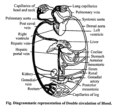
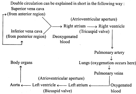
Double circulation: Circulation in which blood flows twice through the heart is called as double circulation. At first heart receives deoxygenated blood from vena cava then send the blood to the lungs for purification through pulmonary arteries. After purification blood is received by heart again through pulmonary vein and then through aorta oxy-genated blood is distributed to whole parts of the body. In double circulation two circulations are found :
- Systemic circulation,
- Pulmonary circulation.
1.Systemic circulation: Vertebrates have a closed blood vascular system. The continuous flow of blood within the system is maintained by the regular rhythmic pumping action of heart. The left ventricle ejects blood into the aorta. Aorta through its branches supply blood to various organs of the body. After the exchange of gases the deoxygenated blood returns to the heart through a large number venules, veins and ultimately to the superior and inferior vena cava. This circulation is known as systemic circulation.
2. Pulmonary circulation: The right ventricle on contraction pumps blood into the pulmonary artery which carries the deoxygenated blood to the lungs. After the oxygenation process blood returns to the left atrium through two pairs of pulmonary veins. This circulation is known as pulmonary circulation.
Question 11.
Describe various heart diseases of man in brief.
Answer:
Heart diseases in man: General symptoms of heart diseases include breathlessness, ankle swelling, liver congestion, dry cough, fatigue and difficulty in lying down. The diseases are described below :
- Arteriosclerosis: In this disease arteries and arterioles lose their elasticity. This loss of elasticity is due to deposition of cholesterol or calcium and thickening of the fibrous tissue in the inner lining of blood vessels. As a result the lumen of the arteries becomes narrow. High blood pressure and cerebral haemorrhage are the common disease symptoms.
- Coronary heart diseases : A set of coronary arteries supply blood to the heart muscle. When they become hard and narrow, blood supply to heart is reduced. The affected person feels severe pain in the chest along the arms. Sometimes a clot may develop and stop the blood supply to the heart. This is called coronary thrombosis. Nitroglycerine tablets relax the arteries and thus reduce the pain. Heparin and dicumarol prevent blood clotting and promote its smooth flow.
3. Rheumatic heart: The disease is caused by the inflammation of the heart. It may affect:
- Inner lining of the heart including valves: It is called endocarditis.
- Outer covering of the heart: It is called pericarditis.
- Heart muscles: It is called myocarditis.
Inflammation may cause permanent damage to heart. Usually, it is followed by the’ infection of streptococci bacteria. The bacteria release the toxin which travels to the joints to cause rheumatic fever, i.e. swelling and pain in legs and fever.
Rheumatism causes permanent damage to heart in two ways :
- Opening of the valves is narrowed and
- Valves do not close properly. Both of these defects hinder the free flow of blood in the required direction.
- Low blood pressure, joints swelling and pain in legs and fever are the typical symptoms. Bed rest is advised.
4. Hypertension: When a person suffers from nervous tension or emotional stress such as fear, worries, anxieties etc. they show high blood pressure because the walls of their arteries get contracted. This is hypertension. If such a stress is frequently found then it results in a persistent high blood pressure.
During this phase heart has to work harder to pump blood to different parts of the body. This may sometimes lead to destruction of the arteries of kidney causing chronic nephritis. Headache, dizziness, fatigue, restlessness are the typical symptoms of hypertension. Chlorothiazides are given as medicines. Bed rest, exercise and avoidance of alcoholism and smoking are advised.
5. Ventricular fibrillation: In this disease each part of ventricles contracts at different time.
6. Angioma: Due to formation of blood clot in the coronary artery, heart wall do not get sufficient supply of blood and causes severe chest pain.
7. Myocardial infraction or Heart attack: Due to obstruction in the coronary artery heart muscle do not get sufficient blood due to which the cells damage and fails to function properly. This condition is called as myocardial infarction or heart attack.
![]()
Body Fluids and Circulation Class 11 Important Questions Objective Type
1. Choose the correct answers:
Question 1.
S. A. node is found in :
(a) Alimentary canal
(b) Aorta
(c) Liver
(d) Heart.
Answer:
(d) Heart.
Question 2.
Sphygmomanometer is used for the measurement of:
(a) Blood pressure
(b) Rate of heartbeat
(c) Rate of blood flow
(d) Temperature.
Answer:
(a) Blood pressure
Question 3.
Blood platelets are concerned with :
(a) Transport of CO2
(b) Release of antitoxins
(c) Production of antibodies
(d) Release of thromboplastin.
Answer:
(d) Release of thromboplastin.
Question 4.
In which form the CO2 is carried out by the blood :
(a) Sodium carbonate
(b) Sodium bicarbonate
(c) Potassium carbonate
(d) Magnesium bicarbonate.
Answer:
(b) Sodium bicarbonate
![]()
Question 5.
An antibody is :
(a) A molecule that specifically inactivates an antigen
(b) W.B.Cs. which invade bacteria
(c) Secretion of mammalian R.B.Cs.
(d) Component of blood.
Answer:
(a) A molecule that specifically inactivates an antigen
Question 6.
Carotid artery carries :
(a) Impure blood from brain
(b) Oxygenated blood to anterior region of body or to brain
(c) Impure blood to kidney
(d) Oxygenated blood to heart.
Answer:
(b) Oxygenated blood to anterior region of body or to brain
Question 7.
During diastole :
(a) Blood enters lungs
(b) Blood leaves the ventricle
(c) Blood leaves the heart
(d) Blood enters the heart.
Answer:
(d) Blood enters the heart.
Question 8.
Heart beat is regulated by the cranial nerve :
(a) Xth
(b) IXth
(c) find
(d) Vth.
Answer:
Question 9.
What is blood bank in human body :
(a) Spleen
(b) Lungs
(c) Heart
(d) Liver.
Answer:
(a) Spleen
Question 10.
Average cardiac output is :
(a) 4 litre per minute
(b) 63 litre per minute
(c) 5-3 litre per minute
(d) 73 litre per minute.
Answer:
(c) 5-3 litre per minute
![]()
Question 11.
Rh factor is a protein present:
(a) On R.B.Cs.
(b) In plasma
(c) On W.B.Cs.
(d) In serum.
Answer:
(a) On R.B.Cs.
Question 12.
During systole of ventricle:
(a) Blood enters the heart
(b) Blood leaves the heart
(c) Blood leaves the ventricle
(d) Blood enters lungs.
Answer:
(c) Blood leaves the ventricle
Question 13.
Pure blood (Oxygenated) is formed in:
(a) Pulmonary vein
(b) Renal vein
(c) Hepatic portal vein
(d) Pulmonary artery.
Answer:
(a) Pulmonary vein
Question 14.
The life of R.B.Cs. is:
(a) About 120 days
(b) 90 days
(c) 30 days
(d) 10 years.
Answer:
(a) About 120 days
Question 15.
Where is chordae tendineare found in the heart:
(a) Ventricle
(b) Left Ventricle
(c) Right Auricle
(d) None.
Answer:
(a) Ventricle
Question 16.
William Harvey is famous for the discovery of :
(a) Respiration
(b) Blood contraction
(c) Blood circulation
(d) digestion.
Answer:
(c) Blood circulation
Question 17.
Which part of vertebrate is responsible for pure blood:
(a) Gill
(b) Lungs
(c) Spleen
(d) Liver.
Answer:
(c) Spleen
![]()
Question 18.
Spleen is:
(a) Haemopoitic
(b) Lymphide
(c) Reproductive
(d) None.
Answer:
(b) Lymphide
Question 19.
Which structure of human when not working properly is replaced by pacemaker:
(a) S. A. node
(b) A.V. node
(c) Both (a) and (b)
(d) None.
Answer:
(a) S. A. node
Question 20.
Which instrument is used for measuring blood pressure :
(a) Stethoscope
(b) Electrocardiograph
(c) Sphygmomanometer
(d) None.
Answer:
(c) Sphygmomanometer
Question 21.
Which vitamin help for clotting of blood :
(a) Vitamin-E
(b) Vitamin-K
(c) Vitamin-C
(d) Vitamin-D.
Answer:
(a) Vitamin-E
Question 22.
Which blood cells kills pathogenic germs in the blood :
(a) Platelets
(b) Red blood corpuscles
(c) White blood corpuscles
(d) Skin cells.
Answer:
(c) White blood corpuscles
![]()
Question 23.
Hormone which regulate heartbeat and blood pressure :
(a) Thyroxin
(b) Adrenalin
(c) Gastrin
(d) Secretin.
Answer:
(b) Adrenalin
Question 24.
Where sinu-auricular node is located :
(a) In the Brain
(b) In the Liver
(c) In the Spleen
(d) In the Heart.
Answer:
(d) In the Heart.
2. Fill in the blanks:
1. The number of chambers in the heart of amphibian is ……………………………………. whereas number of chambers in human heart is ……………………………………. .
Answer:
Three, four,
2. ……………………………………. vascular system is found in sponges.
Answer:
Water,
3. Neurogenic heart is found in ……………………………………. .
Answer:
Invertebrates,
4. The oxygenated blood from lungs reaches left atrium of heart through ……………………………………. .
Answer:
Pulmonary artery,
5. Formation of blood is called ……………………………………. .
Answer:
Haemopoisis,
6. Pericardium is ……………………………………. layered.
Answer:
Two,
7. ……………………………………. is a lymphatic organ.
Answer:
7. Spleen,
8. ……………………………………. heart is found in mammals.
Answer:
8. Myogenic,
9. Blood cells are produced in the ……………………………………. .
Answer:
Bone marrow,
10. ……………………………………. is not found in human RBCs.
Answer:
Nucleus.
3. Match the following:
(A)
| Column ‘A’ | Column ‘B’ |
| 1. AV node | (a) AV. node |
| 2. SA node | (b) Systematic circulation |
| 3. Vena cava | (c) A.V. Bundle |
| 4. Second cardiac sound | (d) Pacemaker |
| 5. First cardiac sound | (e) Semiluner valve. |
Answer:
1. (c) A.V. Bundle,
2. (d) Pacemaker
3. (b) Systematic circulation
4. (e) Semiluner valve.
5. (a) AV. node.
![]()
(B)
| Column ‘A’ | Column ‘B |
| 1. Avian heart | (a) Nerve fibre |
| 2. Reptilian heart | (b) Spleen |
| 3. Purkinje fibre | (c) Left atrium and left ventrium |
| 4. Blood bank | (d) Three chambered |
| 5. Bicuspid valve | (e) Four chambered. |
Answer:
1. (e) Four chambered.
2. (d) Three chambered
3. (a) Nerve fibre
4. (b) Spleen
5. (c) Left atrium and left ventrium.
(C)
| Column ‘A’ | Column ‘B’ |
| 1. Eosinophils | (a) Blood clotting |
| 2. Red blood corpuscles | (b) Universal recipient |
| 3. ‘AB’Bloodgroup | (c) Prevent infection |
| 4. Blood platelets | (d) Contraction of heart |
| 5. Systole | (e) Transport of respiratory gas. |
Answer:
1. (c) Prevent infection,
2. (e) Transport of respiratory gas.
3. (b) Universal recipient
4. (a) Blood clotting
5. (d) Contraction of heart.
4. Answer in one word:
1. Where does dub and lub sounds are produced in cardiac cycle.
Answer:
When auricle contracts, ventricle relax
2. Name the sounds produced due to following actions:
(a) Closing of atrio ventricular valve and initiation of ventricular contraction.
(b) Closing of semilunar valves and initiation of contraction of ventricle.
Answer:
Lub, dub,
3. What is carbaminohaemoglobin?
Answer:
Haemoglobin + CO2,
4. Write the pulse rate of human.
Answer:
72 times per minute,
5. How many types of circulatory systems are found in animals?
Answer:
Two, Open and closed circulatory system,
6. Name the instrument meant for recording heartbeat?
Answer:
Electrocardiogram (ECG),
7. Where are Purkinje fibres situated?
Answer:
Auricular ventricular node (A.V.Node),
8. What is the blood pressure of a normal adult human being?
Answer:
120/80 mm Hg.,
9. Thrombocytes are present in which part of human body?
Answer:
Blood platelets,
10. Why blood is red in colour?
Answer:
Due to Haemoglobin
11. What is blood pressure of a normal man?
Answer:
120/180.